Absence of fibroin H sequences and a significant divergence in the putative fibroin L homolog in Neomicropteryx cornuta (Micropterigidae) silk
 silk")
Introduction
Silk is a water-insoluble proteinaceous material prominent for its bioadhesive properties, and is produced by various Chelicerata and Hexapoda species1. In insects, silk production has evolved independently at least 23 times2. Particularly, most larvae of Lepidoptera (moths and butterflies) and Trichoptera (caddisflies), constituting members of Amphiesmenoptera, synthesize silk via the salivary glands, known as silk glands (SG)3,4. Aquatic caddisfly larvae spin silk under water to construct capture nets, portable cases, and pupation cocoons. Moths primarily use silk for spinning cocoons and making portable cases and protective shelters.
Silk produced by moths and caddisflies comprises few proteins synthesized in the silk glands and stored in a liquid form, which hardens after spinning5. The primary components of silk filaments, heavy and light fibroin chains (FibH and FibL, respectively), are distributed among moths and caddisflies. These fibroin subunits form an insoluble fiber core that provides mechanical strength to the silk. The fibroin core is coated by several layers of soluble, adhesive proteins that bind the fibers5,6. Regarding the silkworm and honeycomb moth, Galleria mellonella, the adhesive layer can be formed by sericins, which are hydrophilic glue proteins7,8, and caddisflies also incorporate several presumptive adhesive proteins (pseudofibroins or cadhezins)9. The silk of moths and caddisflies also contains several other proteins, such as proteases and antibacterial proteins7,10.
Recent genome sequencing has demonstrated homology between several fibH and fibL genes in terrestrial moths and aquatic caddisflies11,12,13,14. The fibH gene is large, has short, conserved characteristic sequences at both ends, and variable repetitive sequences in its central region. Despite conserved regions at the N- and C- terminus, FibH molecules have diversified considerably in both groups13,15. In moths, FibH molecules are characterized by their hydrophobicity and motifs, which can form beta-sheet structures responsible for mechanical strength. In contrast, the FibH molecules of caddisflies are hydrophilic and larger, and mostly lack the amino acid residues required to form beta-sheet crystallites12. In contrast, the fibL gene is much smaller and does not contain repetitive sequences. The protein products of both the fibroin genes form dimers16.
Although there has been progress in the study of silk proteins, the gene and protein profiles of silk from Micropterigidae, a group that probably split from other Lepidoptera over 300–200 million years ago, remain unidentified17,18. To fill this knowledge gap, we provide evolutionary insights into an important biomaterial. We used advanced omics to identify the silk-related genes and their secretory products in N. cornuta. In addition, we extended our search for fibroin genes to another micropterigid species and two species of Eriocraniidae of the family Eriocranoidea, representing a deep node in the non-ditrysian moth phylogeny. We show that the FibH is absent in the silk of N. cornuta, and that the putative homolog of the fibroin light chain (FibL) is either absent or significantly altered. Furthermore, we show that the remaining silk proteins are able to form a tight cocoon even in the absence of fibroins
Results
Morphology of SG and cocoons
To investigate the arrangement of the silk gland (SG) in N. cornuta, we performed micro-CT imaging (a technique that uses X-rays to view the inside of an organ layer by layer). Our 3D study revealed a lateral looping pattern in SG. In the larval model of N. cornuta, SG extended approximately to the middle of the body, terminating at the posterior level of the third abdominal segment (Fig. 1). The anterior part of SG, corresponding to what is typically known as the anterior silk gland (ASG) in the other moths and caddisflies, extends from the cephalic region to the end of the first thoracic segment (Fig. 1A). Subsequently, there was a distinct thickening of SG, continuing up to the first abdominal segment. This corresponds to the middle silk gland (MSG) in other lepidopterans. There was a thin layer of cells on the SG surface surrounding the lumen with stored silk (Fig. 1D). In the first abdominal segment, the SGs expand further and form two loops: a shorter loop toward the head and a longer loop toward the lateral part of the body (Fig. 1). This region may correspond to the posterior silk gland (PSG) observed in other lepidopterans. This was marked by large secretory cells encircling the spacious lumen (Fig. 1E).
 silk")
A 3D visualization of a final-instar larva of a micropterigid moth, N. cornuta. µ-CT images were optically sliced along the longitudinal axis and modeled with the Imaris software (Oxford Instruments). H – head, T1, T2, T3 – thoracic segments, A1, A2, A3 – first three abdominal segments. SG, indicated in blue, extends only up to the middle of the body. The red lines with the letters b–e correspond to the approximate position of cross sections (B–E). B–E Paraplast transverse sections through the larval body of the 5th instar stained with Masson trichrome stain (arrows show the SG). A highly magnified image of the SG is inserted at the top right of the full-body section. B anterior SG; C anterior middle SG; D middle SG; E posterior SG. Scale bars: A 400 µm; (B–E) 200 μm; inset images, 20 μm.
The silk stored in the SG lumen showed notable color differences when stained using Masson’s trichrome (Fig. 1 B–E). The silk material in the ASG appeared red, whereas that in the MSG and PSG appeared dark blue. These color differences suggest that silk undergoes structural changes as it moves through the SG. Notably, this staining method did not show the expected differentiation between the fibroin core and the coating layers within the lumen (Fig. 1).
The cocoon of N. cornuta (Fig. 2) was oval-shaped and ~4 mm long. It featured a thin, compact wall that formed an almost impermeable envelope. The cocoon’s filaments, ~3 µm wide, were ribbon-like and tended to fuse with the adhesive layer, suggesting a high content of sticky material. Notably, the front part of the cocoon included chimney-shaped pores with a diameter of 40–50 µm (Fig. 2A–C). These perforations likely formed through intentional circular movements of the larval head during spinning, thereby enabling environmental interactions with the pupa.
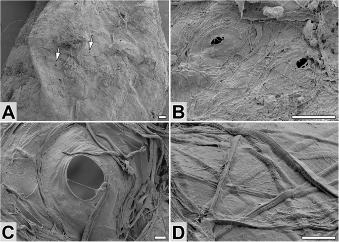
A SEM image of a cocoon with sparse perforations (arrows). B, C Close-ups of cocoon perforation(s). D Arrangement of silk fibers on the outside of the cocoon. Scale bars: (A, B) 100 µm and (C, D) 10 µm.
Detection of putative silk structural proteins
Transcriptome analysis was conducted to identify the major silk components of N. cornuta. First, we isolated RNA and prepared an RNA-seq cDNA library. Transcriptomes of the entire larvae were constructed using de novo and genome-guided transcriptome assembly methods. The de novo transcriptome served as a proxy protein database. Cocoon proteins were digested with trypsin and the resulting peptides were identified using peptide mass fingerprinting by comparing the experimental and predicted MS/MS spectra. We discovered 113 proteins, 62 containing a signal peptide (Table S1). As shown in Table S1, these proteins were categorized into four groups. The first group (13 members) encoded potentially large structural silk proteins characterized by repetitive sequences. The second group comprised 24 members that encoded various enzymes (Table S1). The third group comprised proteins with homologs in other species, but no established association with silk (nine members). The fourth group comprised 16 small proteins with unknown functions, some of which also exhibited repetitive sequences and likely contributed to the structural integrity of silk.
The composition of the N. cornuta cocoon was particularly notable in the first structural group. It comprised gene products organized into small clusters that encode putatively large structural silk components (Table 1). One of these clusters, located in the JAHKQU010000031.1 contig of the publicly available genome ASM2038319v1, included genes encoding sericin-like proteins (Srcl1, Srcl2, and Srcl3). These highly hydrophilic proteins had repetitive sequences with a significant proportion (21–57%) of serine residues. Srcl2 and Srcl3 were particularly large, with molecular weights of 933 and 1450 kDa, respectively. Additionally, four other genes in this group resembled caddisfly cadhezins9. These genes were encoded by two exons and arranged in pairs on two genomic contigs (JAHKQU010000021.1 and JAHKQU010000101.1), with their coding regions split into two exons. Their protein products lacked sequences that were capable of forming crystal domains and did not exhibit sequence conservation with fibroins or sericins. The structural group also comprised four zonadhesin-like genes on the JAHKQU010000031.1 contig that encode protein products between 29 and 130 kDa. These products displayed a high proportion of cysteine residues (14–15%) and conserved Til/EGF2 domains, suggesting their possible role in protease inhibition. Finally, the structural group also included two large genes (containing four and ten exons) encoding putative mucin-like proteins, Muc1 and Muc2, located as singletons on contigs JAHKQU010000032.1 and JAHKQU010000010.1, respectively. These proteins had hydrophilic, repetitive sequences with serine residues comprising 10 and 13% of the sequences, respectively.
N. cornuta silk lacks a close homolog of FibH
Silk proteomic analysis revealed that proteins similar to FibH were absent in N. cornuta. Thus, we performed a comprehensive survey of the transcriptomic and genomic sequences of N. cornuta using the BLAST algorithm, with conserved regions of known fibH genes as reference points, in Lepidoptera and Trichoptera. Our analysis also included the genome sequence of another micropterigid species, Micropterix aruncella. However, we failed to identify fibH-like sequences. These results reinforce the fact that the fibH may have been entirely lost from the genomes of both micropterigid species, or it may have diverged substantially, rendering it unrecognizable by sequence similarity.
The conservation of the genomic region harboring the fibH gene was investigated based on genome sequence analyses of several lepidopteran and trichopteran species, and it was found that the fibH gene is typically located between two genes, prospero (pros) and dihydrolipoyllysine residual succinyltransferase (DRSC). This arrangement is conserved among many lepidopterans and trichopterans, ranging from, Nematopogon swammerdamelus (Adelidae), Incurvaria masculella (Incurvariidae) in Adeloidea, Nymphalis io (Nymphalidae), and B. mori (Bombycidae); additionally, in two caddisflies suborders, including Hydropsyche tenuis (Hydropsychidae) and Parapsyche elsis (Hydropsychidae) in Annulipalpia, and Himalopsyche kuldschensis (Rhyacophilidae) in Rhyacophiloidea. In addition, our study revealed the relatively conserved presence of homologs for the additional two genes, ubiquitously expressed transcript (UXT) and repetitive organellar protein (ROP), in the same genomic region, as shown in Fig. 3.
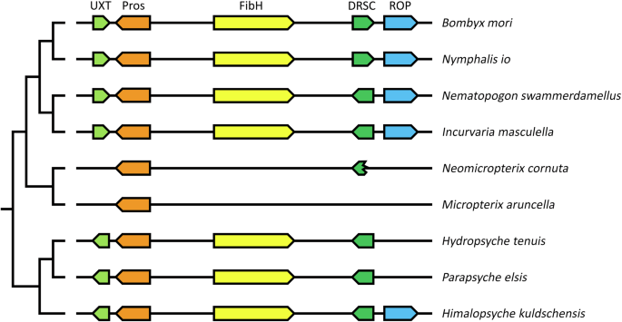
Genes and their relative position and orientation on chromosomes or scaffolds are indicated by block arrows. The same color indicates homologous genes. Homologs of fibH-related genes are shown for four other Lepidoptera and three Trichoptera species. Localization of the region in the represented species: B. mori – chromosome 25 (NC_051382.1); N. io – chromosome 19 (NC_065906.1); N. swammerdamellus – chromosome 20 (OX336353.1); I. masculella – chromosome 19 (OX335659.1); N. cornuta – contig JAHKQU010000018.1; M. aruncella – chromosome Z (OX155980.1); H. tenuis – contig VTON01000064.1; P. elsis – contig JAGVSN010000042.1; H. kuldschensis – contig JAHFWI010000014.1. Abbreviations of the surrounding genes encoding: UXT – Ubiquitously expressed Transcript protein; Pros – Homeobox protein prospero; DRSC – Dihydrolipoyllysine-residue succinyltransferase component of 2-oxoglutarate dehydrogenase complex; ROP – Repetitive organellar protein. B. mori: UXT – XM_062676197.1; Pros – XM_062676169.1; FibH – NM_001113262.1; DRSC – XM_012688929.4; ROP – XM_004921836.5. Nymphalis io: UXT – XM_050498294.1; Pros – XM_050498138.1; FibH – XM_050498483.1; DRSC – XM_050498484.1; ROP – XM_050498112.1.
There were some minor rearrangements within the genomic region, such as the opposite orientation of the UXT gene between caddisflies and moths, an inversion of DRSC in the ditrysian Lepidoptera B. mori and Nymphalis io, and the translocation of ROP in H. tenuis and P. elsis (Fig. 3). Notable conservation of the entire region was observed in most of the Lepidoptera and Trichoptera species that were investigated. Our results also suggest that more extensive restructuring occurred in the two micropterigid genomes, as UXT and ROP relocated to different genomic regions, and the original DRSC copy was lost and replaced by an intronless copy elsewhere in the genome (contig JAHKQU010000032.1, in N. cornuta; chromosome 15 in M. aruncella). Although the 3′-terminal part of the DRSC gene remained at its original position in N. cornuta, the region between Pros and the DRSC residue did not contain any sequence that could encode a larger repetitive protein such as FibH. Overall, fibH was absent in the synteny regions of N. cornuta and M. aruncella; however, it was present in the other species of both groups, supporting the hypothesis that the micropterigids incidentally lose their fibH gene.
Possible N. cornuta ortholog of fibL
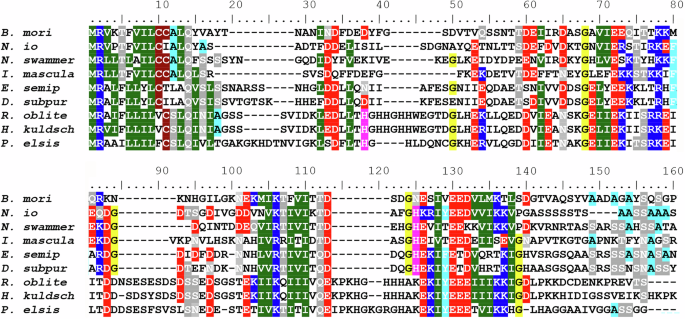
Neither FibH- nor FibL-like proteins were detected in the silk proteins of N. cornuta. To further investigate this, we employed the BLAST algorithm to thoroughly examine the transcriptome and genome sequences of N. cornuta, and the genome sequence of M. aruncella (based on the homology of terminal regions, and location on the contig as opposed to general similarity). We identified a distantly related sequence resembling fibL, named fibX, and organized it into six exons. When comparing the protein encoded by fibX with other FibL proteins from Lepidoptera and Trichoptera, we observed that the similarity was markedly low (Fig. S1). The fibX protein shares only 25–28% identity with other fibL genes. Notably, we did not find any fibL-like gene in the M. aruncella genome.
To assess the conservation of synteny in the genomic regions harboring fibL in Lepidoptera and Trichoptera, we analyzed the corresponding sequences in several representatives of both taxa. We examined the genomes of four moths and one caddisfly (B. mori, I. masculella, M. aruncella, N. cornuta, and Himalopsyche kuldschensis). Our analysis revealed that in the species studied, the fibL gene is on the same contig/chromosome as several conserved genes, including dynein regulatory complex subunit 4 (DRC4) and histone deacetylase 11 (HDAC11). However, the order of these genes is not conserved, preventing us from identifying the exact location of FibL in micropterigids. Notably, the fibX gene in N. cornuta is located on the same contig as DRC4 and HDAC11, suggesting that fibX could be an ortholog of fibL, which has undergone significant divergence. Additional data from micropterigids are needed to confirm that fibX is an ortholog of fibL.
Loss of FibH and the loss or divergence of FibL is specific for Micropterigidae
Is the absence of FibH specific to the family Micropterigidae or does it also apply to other Lepidopterans? For the subsequent analysis, we chose two representative species from the family Eriocraniidae in the suborder Eriocranoidea, which is characterized by its basal split from other Lepidopterans15. We generated transcriptomes of the final-instar larvae of both Eriocraniidae species and performed BLAST searches to identify homologous sequences of fibH and fibL genes. We successfully identified sequences similar to those of fibH and fibL in both eriocraniid species. The resulting alignments of putative FibH and FibL proteins with sequences of several homologs in both Lepidoptera and Trichoptera are shown in Figs. 4 and S1, respectively. Both proteins contain conserved sequences that are characteristic of fibroins. Our results suggest that the absence of fibH and substantial divergence or loss of fibL are specific to Micropterigidae.
The species include seven lepidopterans (Bombyx mori, Nymphalis io, Nematopogon swammerdamellus, Incurvaria masculella, Eriocrania semipurpurella, and Dyseriocrania subpurpurella) and three trichopterans (Hydropsyche tenuis, Parapsyche elsis and Himalopsyche kuldschensis). The accession numbers of the FibH proteins are listed in the legend in Fig. 3; the accession numbers of E. semipurpurella and D. subpurpurella are PQ040461 and PQ040462, respectively.
Discussion
Despite the contrasting habitats of terrestrial moth larvae and aquatic caddisfly larvae, both Lepidoptera and Trichoptera are believed to share a basic silk structure13. Silk in both groups comprises fibroin proteins (FibH and FibL), forming a core surrounded by a heterogeneous group of adhesive coat proteins5. However, various sources of evidence suggest that FibH may have been incidentally lost in the silk produced by micropterigid moth larvae. First, N. cornuta cocoons did not contain any fibroin proteins, as revealed by the proteomic analysis. Second, neither the transcriptome nor the genomic sequence of this species showed sequences closely related to fibH; only one sequence distantly related to fibL was found. Conserved synteny analysis of fibH and the surrounding genes revealed that the region of the fibH gene was absent in the examined micropterigid species. The results of proteomic, transcriptomic, and genomic analyses corroborated the insights from the morpho-histological observations. The liquid silk stored within SG of N. cornuta was only a single layer without distinct core and coat protein layers, and the spun silk was amorphous, resembling that synthesized by the fibroin mutant Nd-s of B. mori (Fig. 5)19,20.
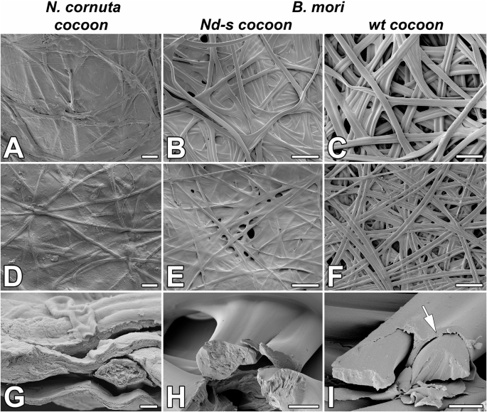
Structure of cocoon and individual silk filaments of N. cornuta A, D, G B. mori mutant in FibL (Nd-s cocoon; B, E, H), and control B. mori (wt cocoon; C, F, I). A–C Scanning electron microscope (SEM) images of the outer side of the cocoon. D–F; SEM images of the inner side of the cocoon. G–I SEM images of broken fibers. Arrows point to the layer of sericin on the surface of B. mori wild-type silk fibers. The structure of the silk at the breaking points shows that the inner part of the silk fiber of N. cornuta is more porous than that of the silkworm. Scale bars: (A, D) 10 µm; (B, C, E, F) 100 µm; (G) 1 µm; (H, I) 10 µm.
The absence of conserved fibH and fibL sequences in micropterigids, coupled with their presence in caddisflies13 and all other examined moths from the suborders Eriocranoidea and Adeloidea, supports the hypothesis that fibH is likely secondarily lost in Micropterigidae while remaining conserved in other Lepidopterans. Consistently, both heavy and light fibroin chains have been previously found in Phymatopus californicus, a member of the family Hepialidae (17), and similar sequences have been also detected in N. swammerdamellus (family Adelidae, https://wellcomeopenresearch.org/articles/8-531). In the two studied micropterigid species, fibL was either absent or highly divergent. The closest sequence identified in N. cornuta, designated fibX, showed only 26.8% identity with fibL from E. semipurpurella. In contrast, the corresponding sequence in the caddisfly P. conspersa showed 39.09% identity with E. semipurpurella fibL. Additionally, a homolog of fibX was not detected in M. aruncella. Phylogenetic analysis indicated that fibX occupied an uncertain position in the cladogram because of its greater divergence (Fig. S2). We propose that FibX diverged after losing its original role as a FibH-binding partner.
These changes in the composition of silk molecules affect the morphology and mechanical properties of cocoons. Species with a higher proportion of fibrillar proteins tend to produce mesh-like silk structures, whereas those with more adhesive proteins form compact continuous layers21. N. cornuta cocoons are compact, smooth, and amorphous (Fig. 5A). These features are comparable to those of G. mellonella cocoons7. Likewise, the fibroin-deficient (naked pupa) mutant of B. mori creates compact, smooth cocoons with tightly bound fibers that are almost devoid of pores20, similar to the dense, sericin-rich cocoons of African moths such as Argema mimosae and Gonometa postica22. This structure, with tiny openings, may facilitate gas exchange or water removal from the cocoon shells and thus may be suitable for the highly humid, typical pupation sites of micropterigid larvae.
Another possible consequence of the loss of fibroin in the micropterigid larvae is the degeneration of spinnerets, a distinctive trait in this family among Amphiesmenoptera (Fig. S3)23,24. Spinnerets are crucial for the final stages of silk formation in silk-producing insects as they apply shear forces essential for converting fibroin into functional silk17,25. Without FibH, the role of the spinneret becomes redundant, potentially leading to gradual degeneration and gradual disappearance.
As the micropterigid moths can still produce cocoons despite lacking fibroins, this challenges the conventional view that the FibH and FibL are highly conserved in the Amphiesmenopteran genomes and play a crucial role in the functionality of their silk. Notably, from the perspective of functional redundancy, they contain several large proteins with putative structural functions, termed as sericin- and cadhezin-like proteins instead of FibH. The amino acid sequences of these proteins are shown in Fig. S4. These proteins have features similar to those of pseudo-fibroins or cadhezins9,10 concerning an exon-intron structure with a typical short first exon and a long second exon with repetitive sequences. Similar to pseudofibroins or cadhezins, N. cornuta cadhezin-like proteins lack the conserved N- and C-terminal regions typical of FibH molecules. In addition, four cadhezin-like proteins, including proline-rich Cazl 3 and 4 and slightly hydrophobic Cazl 1 and 2, may play a structural role. Sericin-like proteins 1, 2, and 3 in N. cornuta are extremely hydrophilic and may be involved in adhesion and/or water uptake. The examined micropterigid species possessed sericin- and cadhezin-like proteins, but their sequences differed significantly. Such differences may be related to the significant evolutionary divergence between these species and the distinct environments wherein these species pupate: under the moss for N. cornuta and in the upper soil layer for M. aureatella26,27. Overall, the presence of unique large silk protein sequences similar to cadhezins and pseudofibroins in aquatic caddisfly larvae may be related to ecological changes in their pupation sites in semi-aquatic environments prone to frequent submersion.
The N. cornuta SG primarily comprises large secretory cells encircling the gland lumen. These secretory cells characteristic of PSG and are specialized to produce large amounts of insoluble fibroin in Lepidoptera and both soluble and insoluble proteins in Trichoptera. In contrast, MSG in moths comprises a thin layer of smaller cells that are primarily responsible for the production of soluble coat proteins. As the MSG in N. cornuta is relatively short when compared to other Lepidoptera species, it can be assumed that the production of coat proteins is limited, as most proteins may be produced in the PSG. Although a thin surface layer is visible in Fig. 6 in the transmission electron microscopy images, we cannot definitively determine the origin of this layer within the MSG.
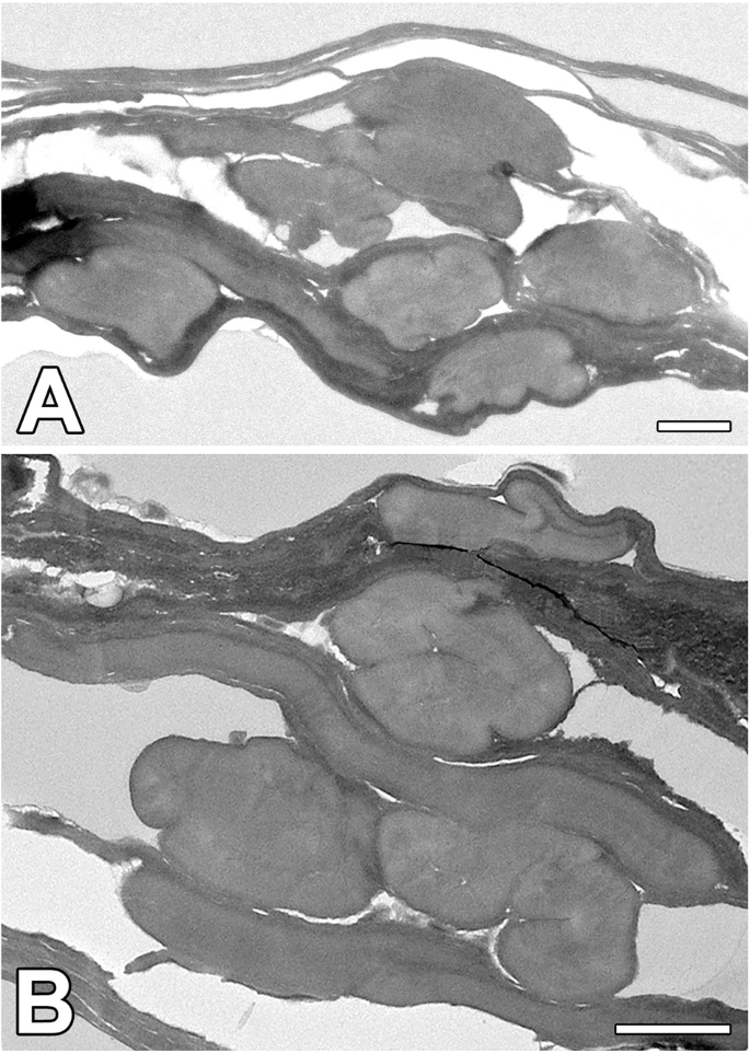
A, B Cross-section through the silk fibers of N. cornuta. Note: the amorphous character of silk fibers. (Scale bars: 1 µm).
As shown in Fig. 5G–I, the silk structure at the breaking points shows that the inner part of the N. cornuta silk fiber is more porous than that of B. mori. Additionally, as shown in Fig. 6, N. cornuta silk exhibits a highly amorphous, putty-like character, which is in contrast with the more rigid structure of wild type and even Nd-s mutant silkworm silk, which retains its shape. We hypothesize that this amorphous nature is advantageous for the formation of cocoons with volcano-shaped openings, as it provides flexibility and adaptability in cocoon construction.
Additional research may uncover other factors contributing to silk protein diversity. Investigating a broader range of species could provide a more comprehensive understanding of the evolutionary divergence and functional redundancy of silk proteins in Amphiesmenoptera. Moreover, functional assays are needed to confirm functional redundancy and structural roles for sericin- and cadhezin-like proteins.
To the best of our knowledge, this study demonstrates the first naturally occurring case of silk production in Lepidopteran species within the Amphiesmenoptera, achieved without FibH and FibL. The proximate cause is secondary fibH gene loss, which is potentially compensated by the function of alternative genes. The unique positioning of Micropterigidae among Amphiesmenoptera in terms of morphology, ecology, and evolutionary history led to this discovery. Exploring the universal functions and redundancy of FibH alongside other proteins and unraveling the adaptive evolution and diversification of these gene clusters and their silk products can provide further insights when compared across diverse lineages representing deep nodes.
Material and Methods
Biological material
N. cornuta final-instar larvae were collected from the mountains of the Shikoku Island at two sites in Tosacho, Kochi Prefecture, Japan. The specimens were pricked with a fine needle and stored in RNA later (Sigma-Aldrich) for several days before RNA isolation. Eriocrania semipurpurella and Dyseriocrania subpurpurella larvae were collected in Ceske Budejovice, Czech Republic.
Micro-CT
Samples were fixed in Bouin-Hollande solution (BHS) for several days at 4 °C without acetic acid, but supplemented with mercuric chloride28 The fixative was then washed in phosphate-buffered saline (PBS). The fixative was then washed in PBS and distilled water and contrasted with Lugol’s solution for 14 days. The samples were scanned with high spatial resolution on a microCT SkyScan model 1272 (Bruker microCT, Land). A total of 1822 projections were acquired with a 360° rotation in 0.1°/ 0.1° rotation steps without filters. The micro-CT scans were performed using the following parameters: Voltage = 40 kV, current = 200 μA, the distance between object and source = 35.2 mm, the distance between detector and source = 275.3 mm, pixel binning = 2 × 2, and total scan duration = 2 h 40 min. Post-processing of the tomographic data: Cross-sectional slices were reconstructed using the volumetric NRecon reconstruction software SkyScan, version 2.2.0.6 (Bruker microCT, Belgium). A 3D model of the SG in the context of the entire larva was created using Imaris software (version 10.0; Oxford Instrument, UK) – Surpass – Contour Surface Module.
Histology and microscopy
Paraffin sections of N. cornuta bodies were labeled with Masson’s trichrome. The cuticles of CO2-anesthetized larvae were punctured with a fine needle using 4% paraformaldehyde as a fixative. Samples were fixed overnight at 4 °C and then rinsed in PBS (thrice for 15 min). Standard histological procedures were used for tissue dehydration, embedding in Paraplast, sectioning (10 μm), deparaffinization, and rehydration. Sections were washed in distilled water and stained with the HT15 Trichrome Staining Kit (Masson) (Sigma-Aldrich, Inc., St. Louis, MO, USA), according to the manufacturer’s protocol. Stained sections were dehydrated and embedded in a DPX-embedding medium (Fluka). High-resolution images of the sections were acquired using a BX63 microscope, DP74 CMOS camera, and CellSens software (Olympus Corporation, Tokyo, Japan) by stitching multiple images and Z-stack imaging.
For transmission electron microscopy: the dehydrated cocoon samples were embedded in resin (Epon) by gradually increasing the volume ratio of resin to acetone (1:2, 1:1, and 2:1, each for 1 h at 20 °C). The samples were incubated in resin for 24 h (20 °C) and then polymerized at 62 °C for 48 h. Ultrathin sections were cut on Leica ultramicrotome, stained with uranyl acetate-lead citrate, and observed using a JEM-1400 JEOL transmission electron microscope (Jeol, Akishima, Japan).
Ultrastructure of silk
Silk samples were cut from cocoons, glued to the surface of aluminum holders, sputter-coated with gold, and analyzed using a JEOL JSM-7401F scanning electron microscope (Jeol, Akishima, Japan).
Transcriptome preparation
Total RNA was isolated from the entire larval body of N. cornuta, E. semipurpurella and D. subpurpurella at the final-instar stage using TRIzol reagent (Invitrogen, Carlsbad, CA, USA)29. RNA sequencing libraries were prepared by NEBNext® Ultra™ II Directional RNA Library Prep Kit for Illumina (NEB #E7765, New England Biolabs, UK). The cDNA library was sequenced on a 2 × 150 bp Illumina platform (paired-end reads) using a NextSeq 500 sequencer. The reads of N. cornuta were mapped to the genome using RNA Star, and the transcriptome was constructed using BRAKER3 (Galaxy Version 3.0.6+ galaxy2). The de novo transcriptome assembly of E. semipurpurella and D. subpurpurella was performed using the Trinity software (v. 2.9.1) on the Galaxy platform30. The de novo and genome-guided transcriptomes resulted in 135,524 and 34,384 assembled contigs, respectively. The quality of the transcriptome assemblies was checked using BUSCO31, using the Insecta odb10 dataset, revealing completeness of 98 and 96%, respectively. The raw data have been deposited in NCBI under the BioProject accession number: PRJNA1215287. Silk gland transcriptome of Neomicropteryx cornuta was deposited in the Dryad repository (https://doi.org/10.5061/dryad.4mw6m90n3).
Protein identification using mass spectrometry
The cocoon proteins were dissolved in 8 M urea and processed according to Hughes et al.32. Briefly, the samples were washed, digested with trypsin, and acidified with trifluoroacetic acid to a final concentration of 1%. The peptides were then desalted using in-house C18 disk-packed tips (Empore, Oxford, USA)33. The processed samples were analyzed by nanoscale liquid chromatography coupled with tandem mass spectrometry (nLC-MS/MS). The resulting MS/MS spectra were compared with the N. cornuta protein database generated from the transcriptome34.
Synteny analysis
For synteny analysis, we used a local BLAST search of the genomic sequences of B. mori, N. swammerdamellus, N. io, I. masculella, H. tenuis, P. elsis, H. kuldschensis, N. cornuta, and M. aruncella using the BioEdit software35. We examined the genomic region surrounding the B. mori fibroin gene on chromosome 25 and identified several conserved genes: Ubiquitously expressed transcript (UXT), prospero (Pros), dihydrolipoyllysine-residue succinyltransferase component of the 2-oxoglutarate dehydrogenase complex (DRSC), and repetitive organellar protein (ROP). The BLAST algorithm was configured to search for homologous sequences with a minimum identity threshold of 80% and a minimum alignment length of 50 base pairs. Plots showing the microsyntenic relationships were then generated based on the best mutual hits between the species.
Phylogenetic analysis
The relationship between the putative fibroin gene from N. cornuta (fibX) and other fibLs genes was also explored using a phylogenetic approach with cDNA sequences from four Lepidoptera and five Trichoptera species. Identities were calculated using the Ident and Sim modules of the Sequence Manipulation Suite36
A basic dendrogram (p-distance, neighbor-joining method) was constructed in MEGA37, whereas a more rigorous phylogram (Maximum Likelihood algorithm) was built using the online version of IQ-Tree38 (http://iqtree.cibiv.univie.ac.at/) with model selected by ModelFinder39 and reliability assessed with ultra-fast bootstrap with 1000 replicates. Graphical editing of the trees was performed in tvBOT40.
Reporting summary
Further information on research design is available in the Nature Portfolio Reporting Summary linked to this article.


Responses