Advanced passive 3D bioelectronics: powerful tool for the cardiac electrophysiology investigation

Introduction
Cardiovascular diseases (CVD) are the leading cause of premature mortality, imposing a substantial burden on population health and socio-economic well-being. The widespread impact of CVD on populations, coupled with the complexity of management, underscores the growing burden on healthcare systems and patients1,2,3. Moreover, drug-induced cardiotoxicity is a significant factor that restricts the development and utilization of new drugs. Many drugs are precluded from clinical application or recalled post-marketing due to adverse cardiac effects. More seriously, drug-induced cardiotoxicity can lead to cardiovascular complications, and in severe cases, may even result in sudden death4,5,6,7. Less than 30% of the compounds tested in U.S. clinical trials successfully reach commercial availability due to drug-induced cardiotoxicity, which leads to a cycle of wasted time, increased expenses, and resource depletion, highlighting the urgent need for accelerated research into all facets of cardiovascular effects8. Electrocardiography, echocardiography, and coronary computed tomography angiography stand as the primary methods to evaluate cardiovascular morphology9,10,11,12. While providing insights into cardiac function, these diagnostic tools are constrained by the low spatial resolution13. To overcome these challenges, cardiomyocyte-based models and sensing technologies are proposed as promising alternatives14,15,16. Establishing a robust cardiomyocyte-based platform is crucial for comprehensive CVD investigation and early anticipation of drug-related cardiotoxicity17.
Electrophysiological sensing of cardiomyocytes is a commonly used and important method for characterizing cell status. Electrophysiological methods are widely recognized for their utility in both characterizing and modulating the behaviors of electrogenic cells18,19. Upon the occurrence of hereditary or acquired CVDs, the generation and propagation of action potentials (AP) are altered due to changes in the expression, distribution, and characteristics of myocardial ion channels20. Thus, evaluating electrophysiological dysfunction at the cell level is crucial to understanding organ dysfunction caused by molecular aberrations21. For electrophysiological evaluation, patch clamp is the gold standard for precise, high-fidelity transmembrane potential measurements, offering detailed, high-resolution insights into ion channel properties, which is labor-intensive and low-throughput22. Automated systems increase efficiency but often require isolated cells, which can disrupt cardiomyocyte electrical coupling and affect measurement accuracy23,24. Extracellular electrodes like planar microelectrode arrays (MEAs) enable noninvasive and easy recording of extracellular APs but cannot capture transmembrane potentials, limiting their ability to reflect detailed arrhythmic features18,25. Alternatively, intracellular recordings from three-dimensional (3D) nanoelectrode arrays (NEAs) can accurately measure AP duration, refractory period, and upstroke velocity, providing precise parameters for electrophysiological properties of cardiomyocytes26,27,28. Therefore, NEAs emerge as highly promising tools in the field of cardiovascular research.
Specifically, there exist two major categories of 3D nanodevices: active and passive. As detailed in Table 1, active devices can actively control or modify electrical signals, and offer capabilities such as amplification, signal fidelity, dynamic modulation, and real-time processing, making them suitable for applications that demand high resolution and precision, particularly in electrophysiology and biomedical sensing. However, active NEAs require an external power source to operate. Passive devices, however, are characterized by their simple structure and independence of an external power source, which facilitates easier and more cost-effective design and manufacturing29. Furthermore, passive devices often exhibit high biocompatibility, generating minimal heat and causing less impact on biological tissues30. Their simplicity also increases stability and durability, with minimal susceptibility to external interference. Additionally, passive devices have low power consumption, making them ideal for miniaturized or implantable applications31. Due to the straightforward design and reduced complexity, passive devices are especially suited for large-scale applications requiring mass production32. Thus, passive devices are widely used for electrophysiological sensing, particularly where active control is not necessary. Despite the aforementioned advantages, the design and 3D structure of the passive devices are crucial for the quality of intracellular signal acquisition.
As for the fabrication of NEAs, micro-nano-processing and integration with 3D bioelectronics allow the recording of electrophysiological signals from both single cells and their networks33. The vertical geometry of NEAs is crucial for strengthening signal detection34. The tight envelopment of the cell membrane around vertical electrodes reduces the separation between the cell membrane and electrode, thereby boosting seal resistance35,36,37. Moreover, for bioelectronic devices to achieve a signal-to-noise ratio (SNR) and amplitude comparable to those in patch clamp, establishing direct contact between recording elements and targeted intracellular locations is necessary while minimizing interference with cell membranes38. To meet these criteria, it is essential to thoroughly understand the size, shape, mechanical properties, and biochemical interactions at the interface between the cell membrane and the nanodevice39. Manufacturing techniques significantly influence nanodevice size, geometry, and the interaction of NEAs with cell membranes. Advances in nanoelectrode technology, particularly through the integration of Complementary Metal Oxide Semiconductor (CMOS) technology, have significantly enhanced the ability to record and modulate cellular electrical activity on a large scale40. Furthermore, by enabling parallel recordings from individual cells and networks, these innovations improve the precision and scalability of electrophysiological studies.
In this review, we will delve into the critical role of arrhythmia as an early diagnostic marker for CVDs, emphasizing the importance of intracellular cardiomyocyte electrophysiological analysis for diagnosis. With the advent of new electrophysiological detection techniques, 3D NEAs have gained prominence for both in vivo and in vitro applications. Here, we focus on the potential of passive 3D NEAs to improve early CVD detection through arrhythmia diagnosis. We discuss the essential aspects that impact electrophysiological recordings using passive 3D NEAs: the manufacturing and configuration of passive 3D NEAs, as well as strategies for intracellular access. We then explore the application of passive 3D NEAs in modeling CVD and precision drug screening. Further, we discuss future research directions in the development of passive 3D NEA structures, including advances in CMOS technology, the integration of micro and nanoelectrodes, multiparameter detection, human organoid models, and the convergence of sensing and regulatory functions.
Manufacturing and configuration of passive 3D NEAs
Manufacturing processes are fundamental in designing and fabricating 3D nanostructures for passive 3D micro-nano bioelectronic platforms and advancing intracellular electrophysiological recordings41. Recent studies have emphasized that the placement, geometry, and dimensions of nanoelectrodes greatly affect electrophysiological detection, with geometry as key to effective cell-device coupling, particularly in 3D configurations42,43,44. Different 3D geometric shapes have been explored for recording electrophysiological signals in cardiomyocytes, each presenting unique innovations, advantages, and challenges38. Here, we provide an in-depth analysis of these strategies to enhance the understanding of nanoelectrode fabrication processes for the precise manufacturing of passive 3D NEAs (Fig. 1).

Physical regulation requires that the electrode penetrates the cell membrane through physical techniques, primarily electro- and opto-poration. Chemical regulation entails gaining intracellular access through chemical methods, such as modifying the electrode surface with a phospholipid bilayer
Advances in passive 3D nanoelectrode fabrication
Since the pioneering work by Thomas et al. in 1972, NEAs have garnered significant attention for the study of excitable cells, particularly cardiomyocytes45,46,47. 3D structures provide significant advantages by enhancing cell adhesion to the nanoelectrode surface, which in turn reduces the electrode’s exposed area and minimizes contact with the surrounding electrolyte solution during recordings. Figure 2 depicts various representative configurations of electrodes as well as the interface between 3D electrodes and cells. To improve coupling between external electrodes and nerve cells by mimicking the neuronal environment, Spira et al. developed gold mushroom-shaped microelectrodes (gMμEs), which typically comprise a stalk, a base, and a cap (Fig. 2a)47,48,49. The fabrication of gMμEs begins by coating silicon or glass samples with a Cr/Au layer using sputtering. Then, samples are spin-coated with photoresistant material. Photolithography, utilizing a photomask, can create openings in the photoresistor. gMμEs are subsequently formed on the surface through gold deposition, before removing the photoresistor layer. The quality of the pattern layer and the stalk thickness are determined by lithography parameters, while the mushroom head size depends on deposition conditions. gMμEs are effective for long-term detection of intracellular APs with improved endocytosis when coated with laminin, allowing for 2–10 min recordings under short electroporation pulses47. However, gMμEs have limitations. One issue is that the success of endocytosis can vary depending on where the cell is located. This variability affects the deformation of the membrane and the quality of the recordings. Additionally, high resistance at the junction between the membrane and electrode can lead to signal loss, which reduces the quality of detection. Solutions to these issues include fabricating a fence structure around the gMμEs to stabilize cell positioning and using low-impedance materials to reduce resistance and improve overall performance48,49. Recent research has demonstrated that vertically aligned nanopillar electrodes can establish strong interactions with cell membranes, significantly reducing impedance by several orders of magnitude through localized electroporation44,50,51,52,53. Nanopillar electrode fabrication involves photolithography for defining Pt tracks and contact pads on quartz substrates, followed by SiO2 deposition via chemical vapor deposition (CVD) under low-pressure conditions for insulation. Reactive ion etching and electron beam lithography create nanoholes above each Pt pad until the Pt layer. Next, Pt nanopillars can be deposited into the nanoholes using a focused ion beam (FIB). Finally, the devices are treated with oxygen plasma for cleaning before coating with a diluted Matrigel matrix and cell plating43. However, nanopillar electrode arrays can also be fabricated through a templated process including atomic layer deposition (ALD), etching, lithography, deposition, and liftoff. The electrodes feature a polyethylene terephthalate (PET) substrate with an Al2O3 core and a Ti/Au conductive coating, insulated with a negative photoresistor layer(Fig. 2b)51. Although nanopillar electrodes offer minimally invasive intracellular recording and are easily scalable, they can suffer from signal loss due to the gap between membrane and electrode. On the other hand, nanowires are ideal as intracellular probes due to their small size, which allows for minimally invasive insertion, while their solid-state nature prevents cell leakage40,54,55,56. Another process involves the fabrication of nanowire electrodes by plasma enhanced CVD (PECVD) for SiO2 layer deposition, stepper lithography, and dry etching to define SiO2 pillars, followed by wet etching to create the nanoelectrode core (Fig. 2c)40. Then, Ti and Pt are sputtered onto the pillars, before adding an ALD passivation layer. A resistor protects the base during wet etching, leaving the metal tip exposed for electrical interrogation. The latest vertical ultrasharp nanowires feature increased height, with tip diameters as small as 10 nm, enabling better cell penetration57. In other cases, iridium oxide (IrOx) nanotubes have been fabricated by cutting a quartz wafer into small pieces, followed by photolithography, Pt/Ti deposition, and liftoff to produce a pattern of electrode pads and lead lines58. The substrate is then coated with Si3N4 and SiO2 layers via PECVD. Plasma etching then exposes the Pt contact pads, and electron beam lithography defines nanoholes on a resist-coated chip. Then, the underlying Si3N4/SiO2 layers are removed to expose the Pt layer, and IrOx nanotubes are electrodeposited into the nanoholes using the resistor as a template (Fig. 2d). The advance of nanotube electrodes leverages cells’ natural propensity to interact with their environment59. In contrast to vertical nanopillar electrodes, the membrane protrusions within nanotube electrodes enhance local membrane tension and reduce the gap between the membrane and electrode60,61. Additionally, ion flows from ion channels or openings within the internal nanotube membrane are more readily detectable before dissipating into the surrounding medium. However, intracellular recordings using IrOx nanotube electrodes suffer from poor SNR and diminished signal amplitude, so addressing these issues is essential for achieving signal quality comparable to that of glass micropipettes and patch clamps60. Similarly, the fabrication of volcanic nanopatterned electrodes begins with successive evaporations of Ti, Pt, Ti, Au, and Ti layers onto a glass wafer, followed by SiO2 sputtering42,62. The substrate is then spin-coated with a photoresistor layer, patterned with 2-μm-diameter openings using a direct laser writer, and Ar+ ion beam etching, followed by photoresistor stripping with O2 plasma42. Electrically conductive tracks are then patterned on a new photoresistor layer, reflowed, placed at an angle, and stripped with O2 plasma. Finally, an insulating SU8 layer is spin-coated to complete electrode fabrication (Fig. 2e). Compared with other nanoelectrodes, nanovolcano (NV) electrodes, with a high aspect ratio and sharp design, offer improved cell access without reducing the effective recording area, leading to lower impedance and higher SNR. Recently, NV electrodes with a large 3D Pt electrode and an insulating SiO2 layer have been developed for stable intracellular recordings lasting >60 min42. Although functionalized gold nanoring was considered critical for sealing, later studies highlighted that tip geometry is more crucial for ensuring a secure cell-electrode interface62. Further research suggests optimizing the shape, size, and materials of NV electrodes, or integrating them with CMOS amplifiers, to enhance recording quality and duration63. Nanocrown electrodes (Fig. 2f) are partially hollow structures featuring an uneven crown edge28,64. The fabrication involves patterning Cr disks via photolithography and using reactive ion etching to form quartz nanostructures64. Ti and Pt are then deposited for metal connections, followed by Si3N4 and SiO2 insulation layers. Afterward, a wet etch reveals the nanoelectrode tips and further etching creates nanocrown structures, completing the fabrication. The nanocrown’s shape has been shown to promote cell membrane wrapping around its surface, improving cell adherence to its core64. This design allows for approximately 60 min of intracellular access to human pluripotent stem cells-derived cardiomyocytes (hPSC-CMs). Additionally, nanocrowns at a depth of 180 nm could record intracellular signals with twice the amplitude compared to those at a depth of 450 nm64.
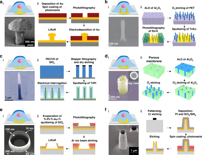
The fabrication processes and representative configurations of the 3D electrode-tissue interface with schematic diagrams and scanning electron microscope (SEM) images of the electrodes. a Mushroom-shaped electrode. Reprinted and modified from Ref. 49 with permission from Frontiers; Ref. 48 with permission from Wiley. b Nanopillar electrode. Reprinted and modified from Ref. 43 with permission from Springer Nature; Ref. 52 with permission from American Chemical Society. c Nanowire electrode. Reprinted and modified from Ref. 40 with permission from Springer Nature. d iridium oxide (IrOx) nanotube electrodes. Inset: Top view showing the hollow center. Reprinted and modified from Ref. 58 with permission from Springer Nature; Ref. 60 with permission from American Chemical Society. e Volcano-shaped electrode. Reprinted and modified from Ref. 42 with permission from the American Chemical Society. f Nanocrown electrode. Inset: Enlarged view of the electrode tip. Reprinted and modified from Ref. 64 with permission from Springer Nature
Innovations in other nanoelectrode designs
Recent advancements in nanoelectrode technology have focused on overcoming the inherent challenges related to the complexity and high costs of conventional manufacturing processes65. A notable innovation was the development of nanobranched electrode arrays, fabricated using a combination of hydrothermal growth and micromachining66. These electrodes are characterized by dense nanobranches with a high aspect ratio, where each electrode measures 20 × 20 μm2. The individual branches have a tip diameter of approximately 200 nm and a root diameter of around 100 nm. This unique configuration enhances the interface between electrode and cell, significantly improving intracellular signal recording. In experimental studies, these nanobranched electrodes successfully recorded intracellular signals from cardiomyocytes for up to 105 min, demonstrating effectiveness in long-term cell recordings66. This capability makes nanobranched electrodes highly valuable for extended electrophysiological studies.
Another significant advancement was the fabrication of nanotrap matrix electrodes from porous PET membranes using microfabrication lithography and magnetron sputtering67. These nanotrap electrodes incorporate traps of varying sizes and densities, strategically designed to promote a tight seal between the cell membrane and the electrode. This tight seal enables self-protruding electroporation, improving the quality and stability of intracellular recordings. This design can extend recording times to approximately 100 min, further demonstrating the efficacy of advanced electrode configurations67.
In addition to these advanced designs, supramolecular self-assembly electrodes have emerged as a promising way to simplify fabrication and reduce the associated costs68. Unlike traditional 3D micro/nanoelectrodes that require complex micro/nano fabrication or solvothermal synthesis, supramolecular self-assembly relies on small organic molecules that efficiently self-assemble into various 3D nanostructures, such as lamellated nanosheets, thin nanobelts, and rod-like structures68. These structures significantly enhance the cell-electrode interface, increasing quality and prolonging intracellular recordings compared to conventional electrodes. The simpler, cost-effective fabrication process makes these electrodes particularly suitable for large-scale electrophysiological studies. Furthermore, the exploration of additional geometrical designs, such as nanorods, offers potential pathways for further enhancing intracellular recording technologies, ultimately paving the way for more accessible and scalable bioelectronic devices65,69,70.
Advanced materials such as graphene, MoS2, and conductive polymers exhibit exceptional biocompatibility, mainly due to their low cytotoxicity, high surface area, and flexibility, which facilitate efficient interaction and integration with cells71,72. These materials promote cell adhesion and growth while minimizing inflammatory responses, ensuring the long-term stability and functionality of the NEAs. Additionally, surface functionalization with bioactive coatings can further enhance cellular interactions by creating a more favorable microenvironment for cell attachment73. This functionalization makes NEAs ideal for chronic applications in electrophysiology and disease modeling, where sustained compatibility with living cells is crucial.
Strategies for intracellular access
To achieve high-fidelity intracellular signals, the nanodevice probe must penetrate the plasma membrane and establish strong coupling to the membrane. Effective intracellular access should enable high-quality, long-term signal recording while preserving cell viability38,39. Intracellular access methods are broadly categorized into electrode surface modification and physical perforation techniques74. Electrode surface modification, including the use of phospholipids or hydrophobic monolayers, enhances cell adhesion and promotes spontaneous membrane penetration75. In contrast, physical perforation techniques, such as optoporation and electroporation, provide a controlled and precise approach by opening transient pores in the cell membrane.
Electrode surface modification
Spontaneous penetration, driven by chemical processes such as endocytosis and adhesion, occurs naturally with minimal impact on cell viability, allowing cells to securely and intimately adhere to the electrode surface76,77. During this process, the lipid bilayer of the cell membrane undergoes a structural rearrangement, forming a tight and robust seal at the penetration site essential for stable intracellular recordings over time. Surface modifications, such as the application of phospholipids or hydrophobic self-assembled monolayers can further optimize this interaction78,79,80. These modifications are specifically designed to enhance the formation of high-resistance membrane seals, thereby improving electrode-cell coupling and promoting more consistent, higher-quality intracellular recordings. Additionally, the precise engineering of these surface coatings improves control over the spontaneous penetration of the electrodes into cells, further reducing signal variability and enhancing the stability and duration of the recorded intracellular signals42.
For instance, hexanethiol-coated electrodes have shown remarkable potential by enabling spontaneous fusion with cell membranes, which supports long-term intracellular recordings (Fig. 3a)42. Although this spontaneous fusion significantly enhances recording duration, key challenges such as the complexity and labor-intensive nature of the modification procedures can make scaling up difficult81,82. Additionally, inefficiencies in consistently achieving reliable intracellular access further complicate the use of such electrodes. Spontaneous perforations, while beneficial, are often infrequent and short-lived, leading to difficulties in maintaining consistent and stable recordings83. These limitations highlight the need for further optimization of the electrode surface to improve both effectiveness and durability. Addressing these challenges is essential not only for enhancing the reliability of intracellular recordings but also for expanding their application potential to large-scale studies and broader research settings38.
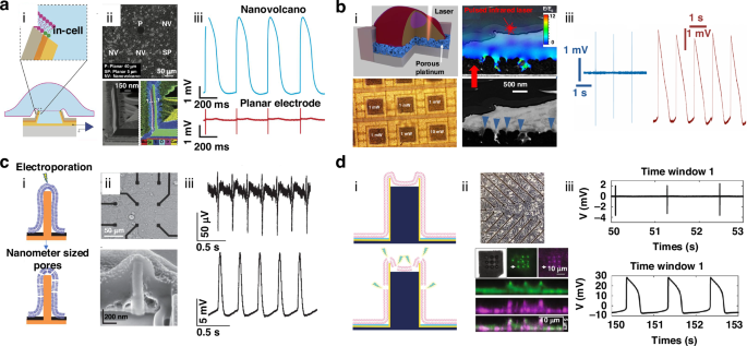
a (i) Chemical-induced spontaneous penetration indicated via a schematic of the Nanovolcano (NV)-cell interface, with dashed lines indicating the cell membrane at the interface where the cell contacts the electrode. (ii) Top, phase contrast image of a typical 3-day-old primary rat cardiomyocyte monolayer cultured on the NV array. Bottom, transmission electron microscopy (TEM) image alongside an energy-dispersive X-ray spectroscopy map of the redeposited multilayered wall. (iii) Extracellular and intracellular recordings. Reprinted and modified from Ref. 42 with permission from the American Chemical Society. b (i) Top, optoacoustic poration by planar meta-electrodes. Bottom, optical imaging of cells on a complementary metal oxide semiconductor (CMOS)-microelectrode array (MEA) after plasmonic optoacoustic poration. (ii) Bottom, SEM images showing cross-sections of HL-1 cells cultured and fixed on the meta-electrodes of a CMOS-MEA, blue arrows indicate the cell’s tightest adhesion points. Top, overlay with a model of the electric field distribution under laser exposure. (iii) Recordings of extracellular and intracellular signals before and after optoacoustic poration. Reprinted and modified from Ref. 84 with permission from Springer Nature. c (i) Electroporation facilitated by a nanopillar electrode. (ii) Top, brightfield images of HL-1 cells grown on a 3 × 3 grid of Pt pads. Bottom, interface between the cell and nanopillar electrode is uncovered through focused ion beam (FIB) milling, showing that the cell fully encapsulates the nanopillar electrode. (iii) Action potential (AP) recordings before and after electroporation. Reprinted and modified from Ref. 44 with permission from Springer Nature. d (i) Electroporation facilitated by nanocrown electrodes. (ii) Top, brightfield image showing a cardiomyocyte monolayer placed on a nanoelectrode array (NEA) device. Bottom, magnified view, enlarged view, and z-projection of a cell positioned on nanocrown electrodes, highlighting the membrane in green and integrins in purple across the vertical span of the nanocrown. (iii) AP recordings before and after electroporation. Reprinted and modified from Ref. 64 with permission from Springer Nature
Physical perforation techniques
Physical perforation methods like optoporation offer significant advantages over chemically induced techniques, particularly in terms of precision and reduced cellular disruption84,85. Optoporation utilizes a focused laser pulse targeted at the electrode-cell interface, inducing various effects such as hot electron injection, thermal heating, and even bubble explosions, all of which facilitate the opening of transient pores in the cell membrane86,87,88. These methods, including plasmonic optoporation and optoacoustic poration, are designed to minimize disturbances to normal cellular activities, thereby supporting stable, long-term intracellular recordings. For instance, Dipalo et al. employed plasmonic optoporation together with vertical nanoelectrodes to generate transient nanopores, allowing uninterrupted intracellular recordings for up to 80 min (Fig. 3b)84. In another example, photoacoustic perforation by mechanical waves from optical pulses together with 3D fuzzy graphene electrodes allowed successful recording of intracellular electrical activity for 20 min85. Both techniques demonstrate a more controlled, less invasive approach to intracellular access, enhancing recording quality and duration without significantly compromising cell viability. As such, optoporation methods show great promise in the development of reliable, long-term intracellular recording platforms, contributing to advanced electrophysiological research and related applications89. However, achieving high-throughput parallel regulation using optoporation methods remains challenging. Integrating a 3D moving platform under the microscope provides a viable solution for enabling multisite optoporation, facilitating precise, high-throughput control of cells.
Electroporation, a widely used technique for intracellular recordings, involves applying electrical pulses to the cellular membrane, creating transient and localized pores that enable extracellular electrodes to capture APs90. Bio-membrane perforation in response to electrical stimuli was first documented in 1972; since then, electroporation has been a leading method in nanoelectrode research due to its simplicity, efficiency, and cost-effectiveness45,91. Electroporation is particularly valued for its ability to achieve intracellular access with minimal complexity. For instance, applying biphasic electrical pulses to HL-1 cardiac muscle cells cultured on nanopillar electrodes enables 10 min intracellular AP recordings (Fig. 3c)44. In a more refined approach, Jahed et al. developed a hollow-nanocrown electrode, which not only enhanced electroporation but also achieved a remarkable 99% success rate, significantly extending both the duration and amplitude of intracellular recordings64. Building on this progress, Fang et al. employed a scalable electroporation strategy using hollow nanopillar electrodes, extending intracellular AP recording durations to 100 min70. In addition, the concurrent recording of both intracellular and extracellular APs using 3D NEAs via electroporation has gained considerable attention. For example, Hu et al. successfully recorded intracellular and extracellular signals from neonatal rat cardiomyocytes using 3D nanobranched electrodes66. While electroporation substantially enhances the amplitude and quality of intracellular signals, it has some limitations. The process may temporarily disrupt the cell’s electrophysiological functions and the duration of intracellular access is constrained by the cell membrane’s natural repair mechanisms, which try to quickly reseal the transient pores created during the procedure62,92,93. Despite these challenges, electroporation remains a powerful tool in electrophysiological research, continuously contributing to the development of more effective recording technologies.
Applications of 3D nanodevices for cardiac electrophysiology
Arrhythmias are closely associated with AP function in cardiomyocytes, having a key role in maintaining the heart’s rhythmic contractions94,95. Proper AP generation and propagation depend on the precise regulation of cardiac ion channels; any disruption in this regulation can result in abnormal heart rhythm or arrhythmia96. Thus, a deep understanding of cardiomyocyte APs is essential for both pharmacological testing and disease modeling, as it allows researchers to study how different drugs or genetic conditions might affect cardiac function. Traditional extracellular recordings, while useful, tend to produce low-resolution signals that fail to capture the full complexity of the waveform, often lacking key information on ion channel dynamics69,97. In contrast, intracellular AP recordings from 3D electrodes offer much more detailed data, providing critical insights into the resting membrane potential and specific ion channel activity98,99,100. Thus, passive 3D nanoelectrodes are particularly valuable in advanced cardiology research, enabling higher-resolution recordings crucial for accurately assessing cardiomyocyte function and investigating the underlying mechanisms of arrhythmias (Table 2).
Cardiomyocyte-based passive 3D nanodevices for drug screening
Recent advances in drug development have increased the prevalence of cardiotoxicity-related issues, leading to drug withdrawal and failures in clinical trials101,102. Certain classes of drugs, particularly anticancer treatments, and antibiotics such as propafenone, flecainide, and terbutaline, have been closely associated with severe arrhythmias due to their significant impacts on cardiac ion channels103. Drug-induced cardiotoxicity can be acute and/or chronic, posing considerable challenges to long-term patient safety and requiring more reliable and efficient cardiotoxicity evaluation methods102,104. As a result, interest in the use of NEA platforms for in vitro drug screening has risen, as these platforms enable the detection of subtle changes in cardiomyocyte APs and ion channel activity in response to drug exposure (Fig. 4)18,98,105,106,107.
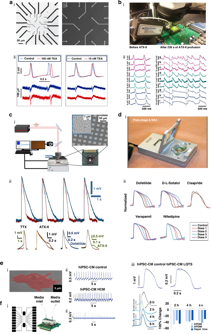
a–d Applications of 3D NEAs for drug screening. a Intracellular APs recorded by nanopillar electrodes after administration of nifedipine and TEA. Reprinted and modified from Ref. 44 with permission from Springer Nature. b Intracellular APs recorded using CMOS-NEA before and after ATX-II administration. Reprinted and modified from Ref. 40 with permission from Springer Nature. c Large-scale optoacoustic poration and intracellular recording in cell networks. Reprinted and modified from Ref. 84 with permission from Springer Nature. d Nanocrown NEA device and intracellular recordings before and after drug administration at varying concentrations. Reprinted and modified from Ref. 64 with permission from Springer Nature. e Intracellular recordings from hiPSC-CMs using nanopillars, showcasing data from a healthy individual and a patient with hypertrophic cardiomyopathy and long QT syndrome. Reprinted and modified from Ref. 43 with permission from Springer Nature. f Intracellular recordings displaying arrhythmic activity following prolonged hypoxic stress. Reprinted and modified from Ref. 114 with permission from the American Chemical Society
Nanodevices employing passive 3D NEAs are emerging as powerful tools in drug screening, providing critical insights into how drugs impact cardiac ion channels and APs108. For instance, Xie et al. utilized nanopillar electrode arrays to investigate the effects of nifedipine, a calcium channel blocker, and tetraethylammonium (TEA), a potassium channel blocker, on APs in HL-1 cells44. A concentration of 100 nM nifedipine caused a significant reduction in APD50 and extended the refractory period, whereas 10 mM TEA had the opposite effect (Fig. 4a). Further, Abbott et al. developed a CMOS-NEA biosensing platform with 1024 channels allowing for extensive and high-resolution intracellular recordings40. After perfusing cells with ATX-II for 239 s, they observed a uniform increase in AP duration across a cellular sheet, with notable regional differences in the extent of APD prolongation (Fig. 4b). Building on this finding, Dipalo et al. integrated CMOS-NEA technology with planar porous electrodes to enhance functionality84. Human-induced pluripotent stem cells were treated with 500 nM dofetilide, 50 μM tetrodotoxin (TTX), and 50 nM ATX-II, which revealed distinct cellular responses, such as slower repolarization (dofetilide), prolonged depolarization (TTX), and altered cellular shapes (ATX-II; Fig. 4c). Moreover, nanocrown electrode arrays demonstrated exceptional sensitivity to subtle drug-induced changes in cardiac electrophysiology, due to fast-sampling rates and self-referential comparison capabilities64. Jahed et al. developed a 57-electrode nanocrown array, which successfully differentiated drug responses, showing that high-risk drugs like dofetilide (0.3 nM, 1 nM, 3 nM, 10 nM) and D,L-Sotalol (0.1 µM, 1 µM, 10 µM, 100 µM) cause a dose-dependent increase in AP duration, while lower-risk drugs such as verapamil (1 nM, 10 nM, 100 nM, 1000 nM) and nifedipine (1 nM, 10 nM, 100 nM, 1000 nM) decreased AP duration (Fig. 4d)64. These advances highlight the potential of passive 3D NEAs in providing detailed and reliable data for drug screening and cardiac safety assessment.
Passive 3D nanodevices for advanced cardiac disease modeling
In vitro modeling has emerged as an essential tool for creating precisely controlled environments that replicate organ and tissue structures with high accuracy109. Advances in cell culture technologies, including cardiomyocyte cultures, stem cell technologies, and 3D tissue engineering, have enabled the cultivation of myocardial tissue that demonstrates synchronized beating, closely mimicking in vivo behaviors110,111,112. When integrated with micro-nano fabrication techniques, biomedical technologies provide researchers with powerful platforms for large-scale investigation of disease mechanisms, offering critical insights to drive significant advancements in the understanding of CVDs113.
Passive 3D NEAs show significant potential for advancing disease modeling, offering more precise and detailed studies of cellular behaviors under pathological conditions. Lin et al. developed an innovative disease modeling platform using a 3 × 3 array of vertical Pt nanopillars to study hPSC-CMs43. The system enabled the simultaneous recording of intracellular APs using nanopillar electrodes together with a conventional patch clamp, providing a more comprehensive view of the cells’ electrical activity. The platform was particularly effective in modeling hypertrophic cardiomyopathy, as it accurately captured arrhythmic activity and delayed depolarization patterns, in contrast to the regular beat intervals observed in healthy cardiomyocytes43. Moreover, the platform successfully demonstrated the prolonged APs characteristic of long QT syndrome, further validating its utility in disease-specific research43. In another study, Liu et al. designed a heart-on-a-chip model to investigate the effects of acute hypoxia on cardiac performance114. This advanced model incorporated a microfluidic channel, a cell culture zone, and embedded electronic components capable of recording both extracellular and intracellular electrical signals. Under hypoxic conditions (1% O2) over a 6-h period, the platform revealed significant decreases in APD50, APD90, and depolarization time in cardiomyocytes, demonstrating the system’s sensitivity to changes in the cellular environment. These effects persisted throughout the study (at 0, 2, 4, and 6 h), providing valuable insights into how hypoxia impacts cardiac electrical activity over time114. Future advances in passive 3D NEAs are expected to involve the integration of CMOS circuits, enabling the development of high-throughput, high-resolution electrophysiological platforms to simultaneously study multiple parameters, offering a more detailed investigation of cellular activities115,116. Additionally, the combination of passive 3D NEAs with tissue engineering technologies holds the potential to create more accurate and biomimetic 3D tissue models for disease studies117,118. Such systems, equipped with multiparameter sensing and regulatory capabilities, could pave the way for comprehensive investigations into the physiological mechanisms that govern cellular functions, ultimately contributing to better understanding and treatment of complex cardiac diseases25,119.
Perspective and conclusion
Emerging trends in micro-nano-bioelectronics, with 3D NEAs at the forefront, are shaping the future of intracellular recordings, particularly in cardiac research. These advanced devices hold tremendous potential for more precise and high-resolution insights into cardiac activity, helping to uncover new therapeutic approaches for heart diseases. However, several challenges must be addressed to fully realize the promise of passive 3D NEAs. First, further optimizing the design and penetration techniques of passive NEAs is critical for achieving efficient interfacing with cardiomyocytes35,108,120. Electroporation faces challenges such as membrane disruption, inconsistent permeabilization, and noise interference during intracellular recordings41. Single-pulse electroporation is considered a less invasive method for the study of cardiomyocytes; however, precise regulation of parameters such as pulse amplitude and duration is critical for enhancing recording fidelity64,121. Advancing single-pulse techniques and integrating advanced noise-reduction strategies are vital for achieving stable, safe, and high-quality intracellular recordings. Second, integration of NEAs with CMOS technology, which has the potential to enable high-throughput, high-resolution intracellular recordings at a large scale122,123,124. CMOS integration significantly improves spatial resolution by minimizing the number of leads at the electrode layer, achieved through shared leads, allowing for thousands of electrodes to be integrated. Leveraging addressable capabilities, these passive NEAs enable recordings at both the single cells and their networks, allowing for precise cell modulation and the detailed mapping of electrical activity propagation within the cells40. Furthermore, combining passive NEAs with CMOS technology boosts scalability by enabling high-density electrode integration, parallel data processing, miniaturization, and cost-effective production, making the system suitable for large-scale applications125. However, while CMOS integration enhances functionality, new technical challenges arise, such as the need for innovative solutions to maintain signal fidelity, manage heat dissipation, and ensure seamless integration of the two technologies123,125,126. Consequently, material selection and structural precision remain critical factors127,128,129. Ongoing research focuses on refining manufacturing methods and material properties to improve the overall performance of these devices38. Compared to traditional inorganic electrodes, new soft and functionalized materials offer significant potential for enhancing the biocompatibility of NEAs. To effectively incorporate these promising biocompatible materials into passive 3D NEAs, developing optimized fabrication techniques that suit their unique properties is essential. Conventional fabrication methods often lack compatibility with soft materials, necessitating the development of innovative dry patterning, low-temperature deposition, and plasma-free processes that are compatible with novel materials130. Certain 2D materials are prone to oxidation in biochemical environments, necessitating the development of surface passivation techniques that not only maintain electrical conductivity but also provide effective insulation against water and oxygen. These advancements would not only improve biocompatibility but also enhance the long-term stability of the devices. Furthermore, future developments in multiparameter detection aim to revolutionize the use of 3D nanoelectrodes, particularly for drug toxicity assessments69,100,131. By enabling simultaneous monitoring of multiple biological parameters, the reliability and effectiveness of the drug screening platforms could be significantly enhanced99,132,133,134. The use of organoids will further strengthen these drug testing systems, providing a more accurate representation of how drugs interact with human tissues135,136,137,138. Beyond drug screening, integrating bioelectronic devices with electrical stimulation technologies offers promising new treatments for cardiac conditions such as arrhythmias139. By applying targeted electrical impulses to heart tissues, these devices could help restore normal heart rhythms140.
In summary, passive 3D NEAs represent promising tools for cardiovascular research and drug development, offering precise, long-term intracellular recordings of cardiomyocytes. Integration with microfluidic systems, CMOS technology, and real-time drug delivery will transform the landscape of CVD research and treatment. In the future, advances in materials, microfluidics, and chip technologies will drive the development of multifunctional, integrated diagnostic and therapeutic platforms for CVDs.

Responses