FBXO22 promotes HCC angiogenesis and metastasis via RPS5/AKT/HIF-1α/VEGF-A signaling axis
Introduction
HCC ranks as the third most common cause of cancer-related fatalities worldwide [1]. This malignancy is characterized by its distinctive vascular nature, featuring an unusually dense vascular network that contributes significantly to its aggressive behavior [2]. Multiple research studies have demonstrated a significant connection between cancer progression, metastasis, and the dysregulation of angiogenesis [3]. Consequently, antiangiogenic therapy has emerged as a promising approach for managing HCC, especially in advanced stages [4]. Given these findings, there is a pressing need for in-depth investigations into the molecular mechanisms underlying angiogenesis and metastasis in HCC.
Multiple studies have demonstrated the involvement of F-box proteins in cancer pathogenesis, influencing a range of physiological and pathological processes such as human cancer cell proliferation, apoptosis, invasion, angiogenesis, and metastasis [5]. Specifically, the FBXO22 protein, belonging to the FBXO family, has been identified as a crucial factor in modulating VEGF-A expression and tumor angiogenesis [6] as well as tumor metastasis [7]. Previous research has demonstrated that FBXO22 acts as an oncogene in HCC, promoting the proliferation of HCC cells [8]. Additionally, angiogenesis is a crucial factor for tumor cell proliferation and metastasis [9, 10]. Thus, our hypothesis is that FBXO22 has a significant impact on both angiogenesis and metastasis in HCC.
Growth factors and cytokines play a vital role in the communication between tumor cells and endothelial cells during tumor angiogenesis [11]. Functional enrichment analysis of differential genes also highlighted a connection between liver malignancy and the VEGF signaling pathway (Fig. S1A). VEGF-A is especially important in this process [12]. Recent research has shown that many cancer cells have the ability to increase VEGF-A expression in order to facilitate tumor angiogenesis, thus suggesting that VEGF-A inhibitors may serve as viable targets for liver cancer treatment [13, 14]. Nevertheless, the precise mechanism through which FBXO22 modulates the expression of VEGF-A remains incompletely elucidated.
This study investigated the mechanisms through which FBXO22 affects angiogenesis and metastasis in HCC. The findings reveal that FBXO22 interacts with RPS5 to enhance tumor angiogenesis and metastasis by modulating RPS5 protein levels via ubiquitination. Additionally, this regulation of ubiquitination results in the activation of the PI3K/AKT signaling pathway, influencing the expression of downstream effectors such as HIF-1α and VEGF-A. Importantly, both in vitro and in vivo experiments demonstrated that FBXO22 depletion enhances the sensitivity of HCC to Lenvatinib. Consequently, this study provides a potential new strategy to improve HCC treatment effectiveness.
Results
FBXO22 expression was frequently elevated in HCC tissues, especially in those with high endothelial vesicle (EV) density, and was correlated with patient survival in HCC
The TCGA and GTEx dataset indicates a significant increase in FBXO22 mRNA levels in HCC tissue compared to normal liver tissue (Fig. 1A). To investigate the role of the FBXO22 gene in HCC development, gene expression analysis was conducted on 366 HCC patients from The Cancer Genome Atlas (TCGA) database. Patients were categorized based on histopathological classification (G3,4 vs G1,2), revealing a positive correlation between FBXO22 expression and the degree of malignant differentiation in HCC (Fig. 1B). Numerous studies have also reported a strong correlation between blood vessel formation and the progression of liver cancer [15, 16], indicating that FBXO22 may play a crucial role in HCC angiogenesis. To further investigate the association between FBXO22 and HCC angiogenesis, an analysis involving 110 tumor samples and adjacent non-tumor tissues from Tongji Hospital was conducted. Additionally, the quantitative real-time polymerase chain reaction (qRT-PCR) and immunohistochemical (IHC) were utilized to evaluate FBXO22 and CD31 (a biomarker for vascular endothelial cells and a marker indicative of EV density) expression. The findings demonstrated a significant elevation in FBXO22 expression in tissues exhibiting a high EV density compared to those with a low EV density (Fig. 1C). Moreover, the expression of FBXO22 was significantly elevated in HCC tissues in comparison to adjacent para-tumor tissues (Fig. 1D). These two trends were confirmed at the mRNA level using qRT-PCR (Fig. 1E, F). Furthermore, a significant positive correlation was identified between the levels of FBXO22 and CD31 mRNA (Fig. 1G). In the Kaplan–Meier and log-rank tests, it was observed that overall survival (OS) was significantly decreased in patients exhibiting elevated levels of FBXO22 (Fig. 1H) and high EV density (Fig. 1I). The survival outcomes for HCC patients with both high FBXO22 expression and EV density groups were particularly unfavorable (Fig. 1J). Table 1 illustrates the correlation between FBXO22 expression levels, EV density, and clinicopathological parameters in HCC patients. Statistical analysis using a Chi-square test revealed significant correlations between FBXO22 levels and EV density with tumor size and vascular invasion. Furthermore, EV density levels were found to be significantly associated with the number of tumors present. Therefore, the expression of FBXO22 is closely related to angiogenesis in HCC patients.
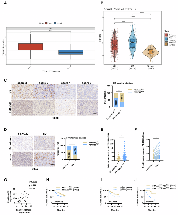
A Comparison of FBXO22 mRNA expression levels between normal liver tissues and HCC tissues in the TCGA database. B The study analyzed the expression distribution of the FBXO22 gene in tumor tissue (grade I/II vs III/IV) and normal tissue. The x-axis represents different sample groups, while the y-axis represents the gene’s expression distribution. Different colors indicate different groups, with significance levels denoted by asterisks in the upper left corner. C Representative IHC images of FBXO22 and CD31 at varying staining intensities in serial sections were utilized to assess EV density in HCC cohort 1 (chi-square test). D The findings indicate that FBXO22 expression is elevated in tumor tissues and is closely associated with CD31 expression levels in HCC cohort 1 (chi-square test). qRT-PCR analysis revealed higher expression levels in HCC tissues compared to adjacent tumor tissues (E) as well as upregulation of FBXO22 expression in HCC tissues exhibiting high levels of CD31 mRNA expression (F). G Correlation analysis further demonstrated a positive association between the mRNA expression levels of FBXO22 and CD31 in 110 HCC tissues. Patients exhibiting high levels of FBXO22 expression (H) and high EV density (I) experienced significantly shorter overall survival (OS). J The log-rank (Mantel–Cox) test further demonstrated that HCC patients with elevated FBXO22 expression and high EV density had significantly poorer OS outcomes (P < 0.0001) when compared to other patient groups. Data are represented as means ± SEM in the bar graphs. *p < 0.05, **p < 0.01, ***p < 0.001, ****p < 0.0001.
FBXO22 promotes tumor cell angiogenesis capabilities in vitro and in vivo
To explore the relationship between FBXO22 and tumor angiogenesis, our research investigated the influence of FBXO22 on the progression of HCC and evaluated its biological roles, including angiogenesis of tumor cells. After analyzing the mRNA levels and protein expression of FBXO22 in six commonly used wild-type liver cancer cell lines (Fig. S1B), stable cell lines were established with either overexpression or knockdown of FBXO22 in specific cell lines. MHCC97H and HLF cells with high endogenous FBXO22 expression were selected for FBXO22 knockdown while HepG2 and Hep3B cells with low endogenous FBXO22 expression were chosen for FBXO22 overexpression. The effectiveness of FBXO22 manipulation was confirmed at both the protein and mRNA levels (Fig. S1C). Based on the correlation identified in liver cancer tissue between the levels of FBXO22 and EV expression, a hypothesis has emerged proposing that FBXO22 could potentially play a role in facilitating angiogenesis within liver cancer. To explore this hypothesis, we collected supernatant from HCC cells with either overexpressed or knocked down FBXO22 and exposed HUVECs (human umbilical vein endothelial cells) to evaluate their impact on angiogenesis-related functions (Fig. 2A) like tube formation, migration, and invasion. These cellular responses serve as indicators of angiogenic potential. The supernatant from cells overexpressing FBXO22 showed increased tube formation, migration, and invasion of HUVECs. In contrast, cells with FBXO22 knockdown exhibited opposite results (Figs. 2B, C and S1D–F). These results suggest that FBXO22 plays an important part in the angiogenesis linked to HCC.
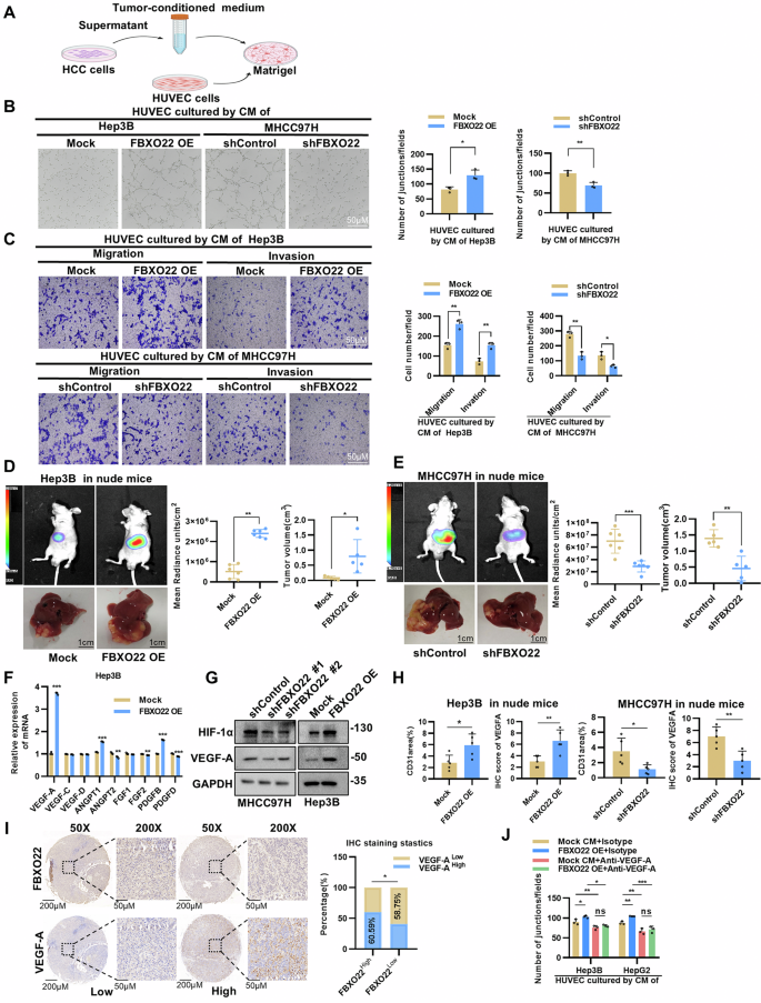
A Tube formation assays were conducted by culturing HUVECs on Matrigel with tumor-conditioned medium (CM), and the tube-forming ability was evaluated by quantifying the number of junctions formed 4–6 h after treatment with CM. B Representative images and the average number of HUVECs junctions cultured by Hep3B FBXO22 overexpression cell line and MHCC97H FBXO22 knockdown cell line. C Representative images and number of migration or invasion HUVECs cells cultured by CM derived from Hep3B FBXO22 overexpression cell line and MHCC97H FBXO22 knockdown cell line. D, E Fluorescence images and a liver diagram (left panel) depicting representative data, along with quantitative results of fluorescence intensity and tumor volume (right panel), were obtained from the Mock, FBXO22OE, shControl and shFBXO22 groups in an intrahepatic model. F Overexpression of FBXO22 resulted in increased gene expression of well-known pro-angiogenic factors, with the most notable upregulation observed in VEGF-A. Total RNA was extracted from Hep3B cells 48 h after plasmid transfection and analyzed using qRT-PCR. G Western blotting showed that downregulation of FBXO22 in MHCC97H cells resulted in decreased HIF-1α and VEGF-A levels. In contrast, upregulation of FBXO22 in Hep3B cells resulted in increased HIF-1α and VEGF-A expression. H Quantitative results of immunohistochemical staining of FBXO22 and angiogenesis-related genes (CD31 and VEGF-A) in intrahepatic tumor models. I The expression of FBXO22 in tumor cells of liver cancer tissue is positively correlated with VEGF-A levels. To assess this relationship, consecutive sections of HCC tissue were embedded in paraffin and subjected to IHC staining for the detection of FBXO22 and VEGF-A expression, along with standard image analysis techniques for patient IHC staining. J The addition of VEGF-A neutralizing antibodies to tumor-conditioned medium inhibited tubule formation in HUVECs induced by overexpression of FBXO22. Conditioned media from Hep3B and HepG2 cells overexpressing FBXO22 were collected and supplemented with 3.2 μg/ml of anti-VEGF-A to induce tubule formation in HUVECs, followed by statistical analysis. Data and error bars are presented as mean ± SD from triplicate independent replicate experiments. The data were analyzed using a paired Student’s t-test for experiments (B, C, D, E, F, H and J). *p < 0.05, **p < 0.01, ***p < 0.001, ****p < 0.0001.
Subsequent in vivo studies validated the tumorigenic impact of FBXO22. When Hep3B cells with FBXO22 overexpression were introduced into the livers of nude mice, there was a noticeable increase in tumor fluorescence intensity and volume compared to the control group (Fig. 2D). On the other hand, MHCC97H cells with FBXO22 knockdown exhibited decreased fluorescence intensity and volume (Fig. 2E). Using qRT-PCR analysis, we initially investigated the correlation between the overexpression of FBXO22 molecules in Hep3B cells and key pro-angiogenic factors such as VEGF, fibroblast growth factor (FGF), angiopoietin (ANGPT), and platelet-derived growth factor (PDGF). The expression of VEGF-A mRNA exhibited the most significant increase (Fig. 2F), and analysis of TCGA data further supported a strong correlation between FBXO22 and VEGF-A in liver cancer (Fig. S2A). Through Western Blot analysis, we confirmed that reducing FBXO22 levels notably decreased the expression of VEGF-A and its upstream transcriptional regulator HIF-1α in MHCC97H and HLF cell lines. Conversely, elevating FBXO22 expression in Hep3B and HepG2 cell lines led to an upregulation of these proteins (Figs. 2G and S2B). Additionally, tumor tissues with FBXO22 overexpression displayed elevated levels of CD31 and VEGF-A staining, whereas tumor tissues with FBXO22 knockdown showed reduced levels of CD31 and VEGF-A staining (Figs. 2H and S2C). The results from the IHC analysis revealed a positive correlation between FBXO22 and VEGF-A expression in human HCC tissues (Fig. 2I). Furthermore, inhibiting VEGF-A in the tumor-conditioned media significantly hindered the tube-forming ability of HUVECs. These HUVECs were cultured with conditioned media from Hep3B and HepG2 cells overexpressing FBXO22 (Figs. 2J and S2D). These results provided convincing evidence for the crucial role of FBXO22 in promoting HCC angiogenesis in vitro and in vivo by mediating the expression of VEGF-A.
FBXO22 enhances the expression of HIF-1α and VEGF-A via the PI3K/AKT signaling pathway to promote angiogenesis and metastasis of tumors
To investigate the role of FBXO22 in the progression of HCC, mRNAs from HLF/Control and HLF/shFBXO22 cells were isolated and subjected to sequencing. This analysis identified 79 differentially expressed genes (DEGs) (fold change >2, p value < 0.05, q value < 0.05) (Fig. 3A). Subsequent Kyoto Encyclopedia of Genes and Genomes (KEGG) pathway enrichment analysis (Table S4) and Gene Ontology (GO) enrichment analysis (Table S5) were conducted to further elucidate the functional implications of these differentially expressed genes. We observed that the PI3K/AKT signaling pathway was significantly represented within the KEGG database (Fig. 3B), while angiogenesis emerged as the top term with the highest enrichment factor in the Gene Ontology (GO) enrichment analysis (Fig. S2E). The significance of the PI3K/AKT signaling pathway in tumor angiogenesis and metastasis has been established in previous studies [17, 18]. Therefore, our investigation focused on examining the phosphorylation status of PI3K/AKT signaling pathway in HCC cells. In HCC cells with FBXO22 overexpression or knockdown, the phosphorylation level of AKT (S473) was significantly increased or decreased, respectively (Figs. 3C and S2F). This is consistent with previous studies [8] indicating that FBXO22 may have a positive impact on the PI3K/AKT signaling pathway, a notion further supported by TCGA analysis (Fig. S2G). Subsequent cytoplasmic and nuclear isolation and extraction experiments revealed that FBXO22 knockdown led to a simultaneous decrease in cytoplasmic and nuclear AKT (S473) phosphorylation (Fig. 3D). The study demonstrated that the increased levels of HIF-1α and VEGF-A caused by FBXO22 overexpression were reduced upon treatment with LY294002, an AKT-specific inhibitor (Figs. 3E and S2H). Similar findings were observed in experiments involving tube formation, cell migration, and invasion using HUVECs (Fig. 3F, G). The results emphasize the role of the AKT signal pathway in cancer-related blood vessel formation. Since the correlation between the expression of PI3K/AKT signaling pathway and VEGF-A in liver cancer and tumor metastasis is well-established [14, 19], this study further investigates the potential role of FBXO22 in promoting tumor metastasis. Interestingly, the findings demonstrate that FBXO22 overexpression promotes migration and invasion in Hep3B and HepG2 cells, as evidenced by increased capabilities in these processes. Conversely, FBXO22 knockdown leads to a significant reduction in migratory and invasive capacities of MHCC97H and HLF cell lines (Fig. S3A–D). Overall, these results suggest that FBXO22 plays a significant role in enhancing the metastasis of HCC through augmentation of tumor cell migration and invasion capabilities. The tail vein lung metastasis model further demonstrated that mice injected with Hep3B cells overexpressing FBXO22 showed an enhancement in tumor lung metastasis, while MHCC97H cells with FBXO22 downregulation led to a suppression of tumor lung metastasis (Fig. S3E). These results from the mouse models provide compelling evidence that FBXO22 is vital in enhancing metastasis in HCC in vivo. Additionally, LY294002 was able to reverse the enhanced migration and invasion capabilities of Hep3B cells induced by FBXO22 (Fig. 3H). In summary, our study shows that FBXO22 boosts the expression of HIF-1α and VEGF-A by triggering the PI3K/AKT signal pathway, thereby promoting tumor angiogenesis and metastasis.
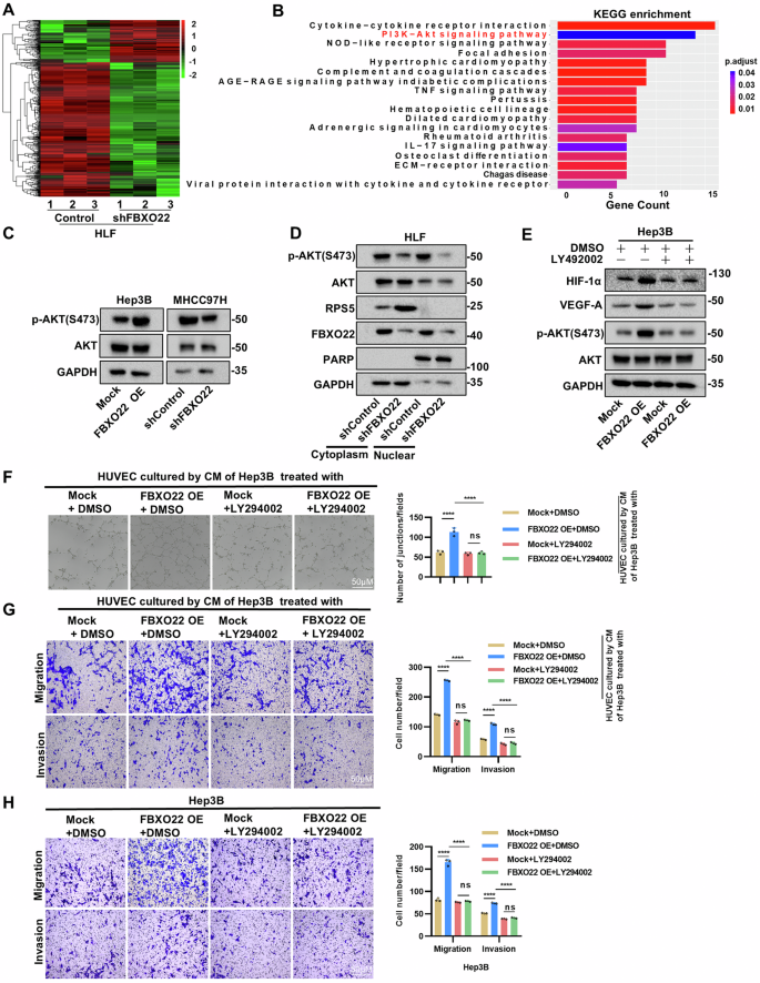
A The heatmap shows the difference between HLF cell control and shFBXO22 expression. B The KEGG analysis of DEGs is displayed. The PI3K-AKT signaling pathway is marked in red. C Overexpression and knockdown of FBXO22 in Hep3B and MHCC97H cells resulted in changes in AKT phosphorylation. D Alterations in the levels of RPS5, AKT, and P-AKT (S473) proteins were observed in both the cytoplasm and nucleus following FBXO22 knockdown. E Treatment with the AKT specific inhibitor (LY294002) (20 μM) reversed the change of HIF-1α and VEGF-A expression, as well as AKT phosphorylation, induced by overexpression and knockdown of FBXO22 in Hep3B cells. LY294002 was administered 24 h after transfection. F, G LY294002 has the ability to inhibit the overexpression of FBXO22, thereby preventing tubular formation and migration and invasion of HUVECs. H LY294002 can prevent the migration and invasion of FBXO22 overexpressed Hep3B cells. Data and error bars are presented as mean ± SD from triplicate independent replicate experiments. The data were analyzed using a paired Student’s t-test for experiments (F–H). *p < 0.05, **p < 0.01, ***p < 0.001, ****p < 0.0001.
FBXO22 interacts with RPS5 and reduces its expression through its ubiquitin ligase activity
To investigate the molecular mechanism by which FBXO22 regulates AKT signaling in liver cancer, we conducted co-immunoprecipitation in HEK293T cells to identify proteins that interact with FBXO22. A total of 77 proteins were found to interact with FBXO22. Using FBXO22 as the bait protein, we selected the top 10 proteins based on spectral count for further verification of their binding to FBXO22 (Table S6 and Fig. S4A). Among these, SNRNP70, RPS5, and PCM1 were identified as interactors of FBXO22 (Fig. S4B). Interestingly, knockdown of FBXO22 did not significantly impact the expression of SNRNP70 and PCM1 (Fig. S4C). However, it was observed that RPS5, a potential interactor, was notably regulated by FBXO22 (Figs. 4A and S4C). In line with the observed interactions during the initial screening, co-immunoprecipitation experiments confirmed the binding of FBXO22 to RPS5 both exogenously and endogenously (Fig. 4B, C). Immunofluorescence staining (IF) demonstrated the colocalization of Flag-tagged FBXO22 and HA-tagged RPS5 in the cytoplasm of MHCC97H and Hep3B cells (Fig. 4D). Functional domains of FBXO22, namely the F-box, FIST-N, and FIST-C domains, were confirmed. Truncation of the FIST-N domain resulted in reduced affinity between FBXO22 and RPS5 (Fig. 4E), indicating the importance of this region for their binding. Moreover, overexpressing FBXO22 in Hep3B and HepG2 cell lines led to a significant decrease in endogenous RPS5 protein expression (Fig. 4F). In MHCC97H and HLF cells, knockdown of FBXO22 resulted in elevated endogenous RPS5 protein levels. Additionally, in order to evaluate potential off-target effects, cells with FBXO22 knockdown were transfected with the FBXO22 plasmid. The subsequent overexpression of FBXO22 in these cells resulted in the reversal of the upregulation of RPS5 (Fig. 4G). In contrast, knocking down or overexpressing RPS5 has no significant effect on the expression of FBXO22 (Fig. S4D). Furthermore, the consistent mRNA levels of RPS5 following both FBXO22 knockdown and overexpression suggest that the regulation of RPS5 may be linked to post-translational modifications (Fig. 4H). This observation motivates further exploration into the potential involvement of ubiquitinating enzyme FBXO22 in the regulation of RPS5. As expected, the study revealed that wild-type (WT) FBXO22, as opposed to inactivated FBXO22ΔF-box, significantly decreased RPS5 protein levels in a dose-dependent manner in HEK293T cells (Fig. S4E). These results suggest that the reduction of RPS5 expression by FBXO22 is reliant on its ubiquitin ligase activity. Subsequently, the investigation explored whether the angiogenesis and metastasis effects induced by FBXO22 are contingent on its ubiquitin ligase activity. Upon re-expression of WT or FBXO22ΔF-box in HLF shFBXO22 cells, it was observed that only the overexpression of WT FBXO22, not the inactivated FBXO22ΔF-box, reinstated the impacts of FBXO22 on RPS5, HIF-1α, VEGF-A, p-AKT(S-473), angiogenesis and metastasis (Fig. S4F–I). These findings further confirm the notion that the oncogenic role of FBXO22 is hinged on its ubiquitin ligase activity.
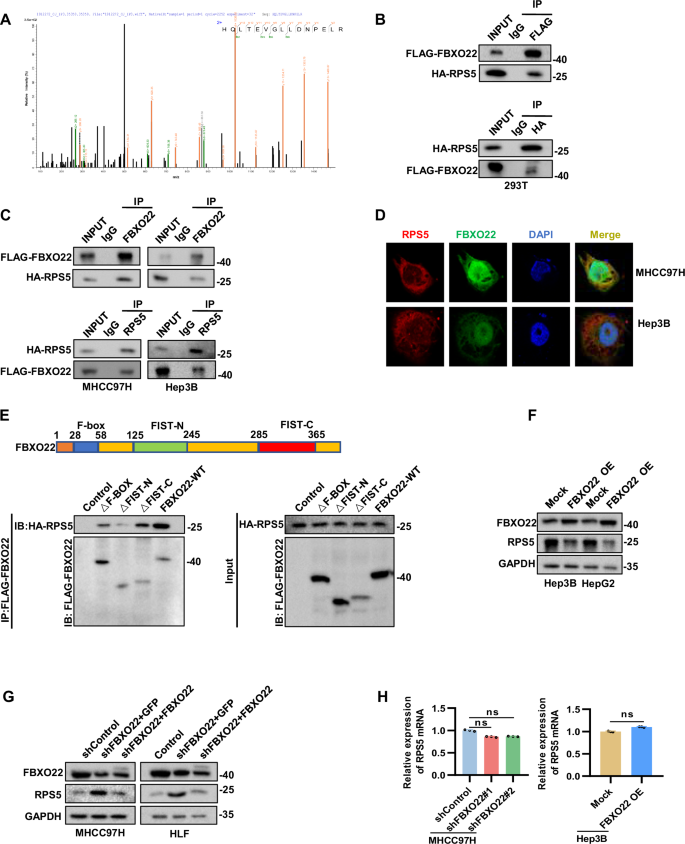
A Mass spectrometry identified RPS5 as an interactor of FBXO22. B, C The interaction was confirmed in HEK293T and HCC cell lines. D Confocal immunofluorescence showed that FBXO22 and RPS5 colocalized in the cytoplasm. E Functional regions of FBXO22 and interactions with RPS5 were shown in a schematic diagram. F Constructs were transfected into Hep3B and HepG2 cells. G FBXO22 knockdown cell lines were transfected with the indicated constructs. H The mRNA expression was detected in the indicated cells. Data and error bars are presented as mean ± SD from triplicate independent replicate experiments. The data were analyzed using a paired Student’s t-test for experiments (H). *p < 0.05, **p < 0.01, ***p < 0.001, ****p < 0.0001.
Ubiquitination-mediated degradation of RPS5 by FBXO22 on Lys85
Our research findings suggest that upregulating FBXO22 expression in Hep3B cells results in a dose-dependent reduction in RPS5 levels. This effect was reversed by the proteasome inhibitor MG132, indicating a potential regulatory role of FBXO22 on RPS5 expression in a proteasome-dependent manner (Fig. 5A). Further investigation into the regulatory role of FBXO22 on RPS5 in a proteasome-dependent manner was conducted in liver cancer cell lines MHCC97H, Hep3B, HLF, and HepG2. Our results showed that treatment with MG132 effectively counteracted the effects of FBXO22 on RPS5 (Figs. 5B and S5A). After treating Hep3B and MHCC97H cells with cycloheximide (CHX) to block protein synthesis, we conducted an analysis of protein extracts at specific time points. Our findings showed that overexpression of FBXO22 in Hep3B cells led to a notable decrease in RPS5 half-life, while knockdown of FBXO22 in MHCC97H cells resulted in a significant increase in the half-life of RPS5. Moreover, there was no significant difference in the half-life between the experimental and control groups following the addition of MG132 (Figs. 5C and S5C). In summary, FBXO22 reduces the expression of RPS5 through proteasome degradation.
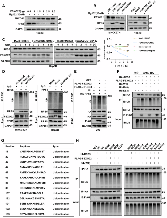
A RPS5 protein levels changed after gradient transfection of FBXO22 plasmids into Hep3B cells. B MHCC97H and Hep3B cells were treated with Mg132 (10 μg/ml) for 8 h, total protein was extracted and analyzed by western blot. C Hep3B cells were treated with CHX (10 μM) and MG132 for specified durations. After treatment, cells were harvested and subjected to immunoblotting for FBXO22, RPS5, and GAPDH. RPS5 levels were quantified relative to GAPDH expression. D MHCC97H and Hep3B cells were treated with MG132 (10 μg/ml) for 8 h, followed by total protein extraction and Western blot analysis. E HEK293T cells were transfected with HA-RPS5, ubiquitin, Flag-FBX022, and Flag-FBX022ΔF-box in combination, followed by treatment with Mg132 (10 μg/ml) for 8 h. total protein was extracted and analyzed by western blot. F Ubiquitination co-IP assay illustrating effect of FBXO22 on K48-linked and K63-linked ubiquitination of RPS5. G Ubiquitination sites of RPS5 predicted by Protein Lysine Modification Database. H Ubiquitination IP analysis showed changes in ubiquitination levels of both wild-type and mutant RPS5 when FBXO22 was present. The lysine residue was mutated to arginine. *p < 0.05, **p < 0.01, ***p < 0.001, ****p < 0.0001.
Considering FBXO22 acts as a ubiquitin ligase and the F-box region is essential for substrate ubiquitination [20, 21], it is hypothesized that FBXO22 reduces RPS5 expression through ubiquitination. Consistent with this hypothesis, our ubiquitination experiments demonstrated that decreased FBXO22 expression in MHCC97H and HLF cells correlated with reduced ubiquitination of RPS5, whereas overexpression of FBXO22 in Hep3B and HepG2 cells led to increased ubiquitination of RPS5 (Figs. 5D, S5B and S5D). In conclusion, FBXO22 mediates RPS5 degradation through a proteasome-dependent mechanism involving ubiquitination. Moreover, transfection experiments were conducted in HEK293T cells with HA-RPS5, ubiquitin, Flag-FBXO22 (wild type), and Flag-FBXO22ΔF-box. Subsequent immunoprecipitation and Western blot analyses revealed a significant increase in polyubiquitinated RPS5 protein levels in cells transfected with Flag-FBXO22 (wild type), while cells transfected with Flag-FBXO22ΔF-box did not show this increase (Figs. 5E and S5E). The findings suggest that FBXO22 predominantly modulates the ubiquitination of RPS5 via its F-box domain. In addition, we co-transfected RPS5, FBXO22, and ubiquitin plasmid into HEK293T cells. The co-immunoprecipitation results indicated that FBXO22 notably increased the polyubiquitination level of RPS5, particularly the K48-linked while showing no significant impact on the K63-linked polyubiquitination level (Fig. 5F).
To identify the specific lysine sites responsible for RPS5 ubiquitination, we examined 13 known lysine ubiquitination sites (K18, K22, K42, K44, K47, K63, K85, K86, K167, K182, K191, K192, K193) on RPS5 from the protein lysine modification database (Fig. 5G). Each of these lysine sites was mutated to arginine. Results from ubiquitination immunoprecipitation (IP) experiments suggest that FBXO22 does not enhance the ubiquitination of RPS5 with a mutation at lysine 85 (K85) (Fig. 5H). In conclusion, FBXO22 targets RPS5 for degradation via the proteasome pathway by boosting K48-linked ubiquitination at the lysine 85 site in the cytoplasm.
FBXO22 interacts with RPS5 to activate the PI3K/AKT signaling pathway, promoting angiogenesis and tumor metastasis
The impact of FBXO22 on the PI3K/AKT signaling pathway was investigated through its role in the ubiquitination of RPS5. Endogenous RPS5 was knocked down in Hep3B cells, while RPS5 was overexpressed in HLF cells (Fig. S6A). In contrast to the impact of FBXO22 on HCC cells, our study revealed that RPS5 decreased HIF-1α, VEGF-A expression, angiogenesis, and metastasis in HCC cells (Figs. S6B–E and S7A–C). Notably, RPS5 also suppressed the phosphorylation of AKT (S473) (Figs. S6B and S6D, E), aligning with findings from previous literature [22]. Depletion of RPS5 in the cytoplasm of liver cancer cell lines HLF and 97H reversed the decrease in AKT (S473) phosphorylation induced by FBXO22 deficiency. This led to elevated expression of VEGF-A and HIF-1α, as demonstrated by cytoplasmic and nuclear isolation and extraction experiments (Figs. 6A and S7D). Knockdown of RPS5 restored tube formation and migration ability in HUVECs cultured in conditioned media from cells with low FBXO22 expression (Figs. 6B, C and S7E). Additionally, knockdown of RPS5 in FBXO22-deficient cells enhanced migration and invasion capacities compared to the control group (Figs. 6D and S7F). Intrahepatic tumor and tail vein metastasis models demonstrated that knockdown of RPS5 in FBXO22 knockdown cells increased tumor burden, CD31, VEGF-A, p-AKT(S473) levels, and lung metastasis in HCC, supported by IHC analysis (Figs. 6E and S7G). These findings suggest that FBXO22 may promote angiogenesis and tumor metastasis in HCC by regulating RPS5 levels and activating the PI3K/AKT signaling pathway.
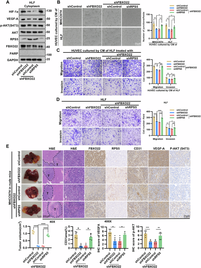
A HLF-shFBXO22 cells were subjected to shRPS5 knockdown, followed by cell nucleocytoplasmic separation experiments. Proteins located in the cytoplasm were isolated, and Western blot analysis was conducted to assess the levels of AKT, phosphorylated AKT (S473), VEGF-A, and HIF-1α in the cytoplasm. B, C The results of HUVECs tubule formation and trans-well assays suggest that the downregulation of RPS5 effectively counteracted the suppressed tubule formation, migration, and invasion of HUVECs when exposed to the conditioned media of FBXO22 knockdown. D Trans-well assays further demonstrated that the suppression of RPS5 resulted in the restoration of migratory and invasive capabilities in HLF cells with FBXO22 knockdown. E Representative images of H&E staining and IHC staining of FBXO22, angiogenic-related genes (CD31 and VEGF-A), and p-AKT(S473) in an intrahepatic tumor model, along with quantitative results of tumor volume, CD31, VEGF-A, and p-AKT(S473). Data and error bars are presented as mean ± SD from triplicate independent replicate experiments. The data were analyzed using a paired Student’s t-test for experiments (B–E). *p < 0.05, **p < 0.01, ***p < 0.001, ****p < 0.0001.
Enhancing Lenvatinib sensitivity in vitro and in vivo by targeting FBXO22
Research has shown that Lenvatinib can target VEGFR and VEGF-A, leading to a reduction in tumor angiogenesis [23]. Additionally, studies have found that downregulation of the PI3K/AKT signaling pathway enhances the therapeutic impact of Lenvatinib [24]. The increased expression of FBXO22 in HCC cells is linked to higher VEGF-A levels and activation of the PI3K/AKT signaling pathway, promoting tumor progression. Therefore, it is suggested that FBXO22 may influence the drug sensitivity of Lenvatinib.
The results of the study demonstrate that decreased levels of FBXO22 resulted in a significant improvement in the in vitro responsiveness of Lenvatinib (Fig. 7A). This improvement in the therapeutic efficacy of Lenvatinib in HCC was achieved by inhibiting the PI3K/AKT signaling pathway and reducing HIF-1α and VEGF-A expression (Fig. 7B). Additionally, the study confirmed that reintroducing FBXO22 into FBXO22-knockdown MHCC97H cells effectively restored tube formation in HUVECs cultured with their conditioned medium, even in the presence of Lenvatinib (Figs. 7C and S8A). Subsequently, these findings were validated in an in vivo model using nude mice. To evaluate the therapeutic impact of FBXO22 on HCC in vivo, adeno-associated virus 8 (AAV8) was used to deliver short hairpin RNA targeting FBXO22 in a murine model. In an intrahepatic tumor model, MHCC97H cells were introduced into the livers of nude mice. Four days later, scAAV8-U6-shNC and scAAV8-U6-shFBXO22 were administered intravenously (Fig. 7D). The successful knockdown of FBXO22 in vivo was confirmed by western blot analysis of mouse liver tissue (Fig. 7E). As expected, the study showed that reducing FBXO22 expression significantly inhibited tumor progression and enhanced the anti-tumor efficacy of Lenvatinib (Fig. 7F), which was further supported by IHC staining and analysis (Fig. 7G). In conclusion, the findings suggest that targeting FBXO22 leads to decreased progression of HCC and improves the therapeutic effectiveness of Lenvatinib by suppressing the PI3K/AKT signaling pathway-mediated expression of HIF-1α and VEGF-A.
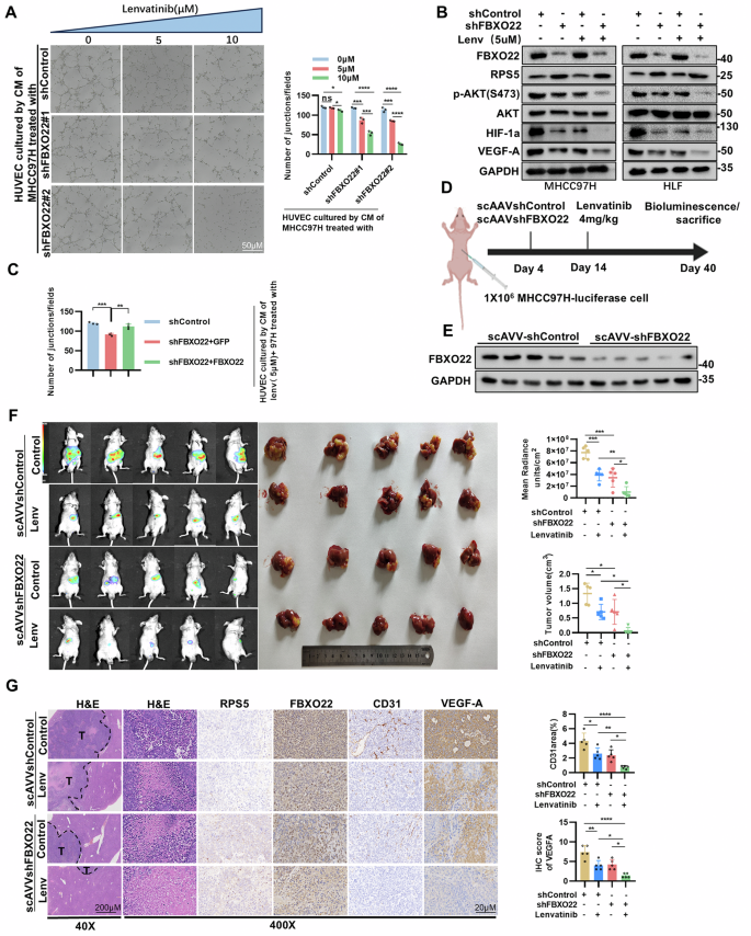
A The effect of Lenvatinib on tubule formation of HUVECs was significantly increased by inhibiting FBXO22 expression in MHCC97H cells (left panel). Quantitative analysis of the effect of FBXO22 on Lenvatinib against tubule formation of HUVECs (right panel). B The suppressive impact of Lenvatinib on the expression of P-AKT(S473), HIF-1α, and VEGF-A were augmented following the knockdown of FBXO22. C Re-expressing FBXO22 could rescue the tubule formation of HUVECs cultured by CM of FBXO22 knockdown MHCC97H cells. D Schematic diagram of AAV8 administration and Lenvatinib gavage in nude mice with orthotopic MHCC97H-luciferase cells. E The expression level of FBXO22 protein in tumor tissue. F The bioluminescence and liver with tumor images of orthotopically injected HCC MHCC97H-luciferase cells in nude mice, following self-complementary AAV8 (scAAV8)-U6-shNC/shFBXO22 administration in combination with Lenvatinib for 40 days. The quantitative results of tumor bioluminescence and volume are shown. G Representative images of H&E and IHC as well as quantitative results for CD31 and VEGF-A from intrahepatic model. Data and error bars are presented as mean ± SD from triplicate independent replicate experiments. The data were analyzed using a paired Student’s t-test for experiments (C, F, G). *p < 0.05, **p < 0.01, ***p < 0.001, ****p < 0.0001.
Correlation between FBXO22 and RPS5 in clinical samples
To investigate the association between FBXO22 and RPS5 in HCC, IHC analysis was performed on a tissue microarray consisting of 110 pairs of liver cancer samples. The results indicated a negative correlation between the expression levels of FBXO22 and RPS5 in the clinical specimens (Fig. 8A, B). Additionally, the protein expression levels of FBXO22 and RPS5 were assessed in 40 pairs of HCC tissues. Western blot analysis revealed a significant increase in FBXO22 expression and a decrease in RPS5 expression in the tumor group compared to the non-tumor liver group (Fig. 8C and Fig. S8B). Correlation analysis showed a negative correlation between FBXO22 and RPS5 (Fig. 8D). These findings suggest an inverse relationship between the levels of FBXO22 and RPS5 in clinical samples, further supporting the mechanism by which FBXO22 may promote HCC progression through the downregulation of RPS5.
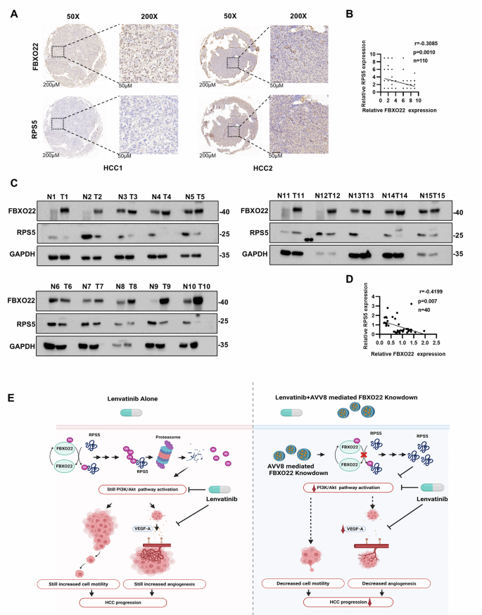
A Representative images of IHC staining with anti-FBXO22 and anti-RPS5. B The expression of FBXO22 was inversely correlated with that of RPS5. C Western blotting to compare FBXO22 and RPS5 expression in liver cancer and healthy tissues. D The expression of FBXO22 was negatively correlated with RPS5. E Integrated model diagram depicts that the FBXO22/RPS5/AKT/HIF-1α/VEGF-A signaling cascade is significantly involved in facilitating angiogenesis, metastasis, and augmenting the efficacy of Lenvatinib in treating HCC.
Discussion
Numerous studies have documented the involvement of FBXO22 in the progression of various cancers [25,26,27], including HCC [8]. In this study, our analysis of public databases suggests a positive correlation between the expression of FBXO22 in liver cancer tissue samples from clinical cases and the degree of tumor malignancy. However, further research is needed to elucidate the exact role of FBXO22 in promoting angiogenesis and metastasis in HCC. This study elucidates the role of the FBXO22/RPS5/AKT/HIF-1α/VEGF-A signaling pathway in facilitating angiogenesis and metastasis in HCC. FBXO22 interacts with RPS5 and regulates RPS5 through the ubiquitination pathway. The downregulation of RPS5 expression through ubiquitination leads to the activation of the PI3K/AKT signaling pathway, subsequently enhancing the expression of downstream effectors HIF-1α and VEGF-A, thereby facilitating tumor angiogenesis, tumor metastasis and sensitivity to Lenvatinib (Fig. 8E).
To date, FBXO22 has been shown to exhibit oncogenic properties through the facilitation of ubiquitination and subsequent degradation of various substrates, such as KDM4A, KDM4B, methylated p53, KLF4, p21, LKB1, CD147, Snail, PTEN, Bach1, and HDM2 [25, 27,28,29,30,31]. The RPS5 molecule was identified through mass spectrometry analysis in the current study as binding to FBXO22. RPS5, a constituent of the 40S ribosomal subunit, plays a crucial role in ribosomal maturation and translational initiation by primarily interacting with initiation factors [32]. The dysregulation of RPS5 has been documented in numerous cancer types [33, 34] and prior research indicates that RPS5 may represent a promising therapeutic target for HCC [35]. Moreover, a separate investigation demonstrated that suppression of RPS5 expression resulted in phosphorylation of AKT at Ser473, thereby inducing activation of the PI3K/AKT signaling pathway and exacerbating liver fibrosis [22]. Our findings indicate that FBXO22 interacts with RPS5 via the F-box domain, leading to the downregulation of RPS5 expression through the ubiquitination pathway.
It is well known that numerous signaling pathways regulate the angiogenesis and metastasis of tumor cells, including the mTOR signaling pathway [36, 37], MAPK signaling pathway [38], and JAK/STAT signaling pathway [39]. However, our research group’s previous knockdown of FBXO22 in HCC, along with the detection of expression changes in key proteins within these signaling pathways [8], as well as the results from transcriptome library sequencing analysis conducted in this experiment, consistently indicate that altering the expression of FBXO22 in HCC significantly impacts the PI3K/AKT signaling pathway. The PI3K/AKT signaling pathway has been identified as a promoter of angiogenesis and metastasis in HCC [40]. FBXO22 has been shown to alleviate rotenone-induced neurotoxicity by promoting the degradation of PHLPP1 via ubiquitination, thereby activating the PI3K/AKT signaling pathway [41]. Nevertheless, limited research has been conducted on the impact of FBXO22 on the PI3K/AKT signaling pathway in cancer. Initially, we assessed the impact of FBXO22 on the processes of angiogenesis and metastasis in HCC using both in vitro and in vivo experimentation. Subsequently, the phosphorylation levels of key signaling regulatory molecules within the PI3K/AKT signaling pathway in HCC cells were determined subsequent to the modulation of FBXO22 expression. Our study further clarified the involvement of FBXO22 in the modulation of angiogenesis and metastasis in HCC through the PI3K/AKT signaling pathway.
Notably, the loss of PTEN protein is closely associated with the PI3K/AKT signaling pathway. Previous studies have shown that FBXO22 molecules can degrade PTEN protein in the nucleus via the ubiquitination pathway in colon cancer cells. However, the study also highlights that degradation of nuclear PTEN by FBXO22 does not further activate AKT and its regulatory effect on PTEN in the cytoplasm is limited [25]. We know that the activation process of PI3K/AKT signaling pathway is closely related to protein molecules in the cytoplasm [42]. Interestingly, we reveal that FBXO22 induces phosphorylation of the AKT (S473) site in both the nucleus and cytoplasm of liver cancer cells. In addition, our findings supported the idea that FBXO22 facilitates the ubiquitination and degradation of RPS5 at Lys85 in the cytoplasm, consequently activating the PI3K/AKT signaling pathway and enhancing angiogenesis and metastasis in HCC. Nevertheless, the specific mechanism of AKT activation in the nucleus and the potential involvement of other molecules in this pathway necessitate further investigation.
The complexity of HCC presents a significant challenge in the development of effective therapeutics [43]. Lenvatinib, as a primary treatment option, has shown promise in improving the prognosis of HCC patients [44]. Nevertheless, a substantial number of patients exhibit either a lack of response or develop resistance to the drug in a short period of time [45]. Hence, it is imperative to investigate the synergistic effects of combining Lenvatinib with other pharmaceutical agents in order to optimize therapeutic outcomes. Our research suggests that targeting the FBXO22/AKT/HIF-1α/VEGF-A axis may serve as a promising strategy for enhancing the efficacy of Lenvatinib. In order to evaluate this hypothesis, the efficacy of HCC cells with FBXO22 knockdown on Lenvatinib was investigated in vitro setting. Due to the lack of inhibitors, we utilized shRNA targeting FBXO22 to inhibit its expression, and conducted in vivo experiments using the adeno-associated virus vector ScAAV8-U6-fbxo22(m)-zsgreen-shRNA for delivery. Our study revealed that the downregulation of FBXO22 resulted in the effective inhibition of HCC tumor growth and significantly improved the therapeutic efficacy of Lenvatinib. This observed regression of the malignant phenotype in HCC following FBXO22 knockdown can be attributed to the suppression of the PI3K/AKT signaling pathway, as well as the notable decrease in HIF-1α and VEGF-A expressions.
FBXO22 plays crucial roles in tumor development by degrading various substrates across different cancers. In HCC [8], lung cancer [31], colorectal cancer [25] and breast cancer [20], FBXO22 exhibits cancer-promoting functions; however, in breast cancer [20], it is suggested that FBXO22 may function both as a tumor suppressor gene and as an oncogene. Consequently, to gain a deeper understanding of the role of FBXO22 in cancer, several fundamental questions remain to be addressed. For instance, what unknown regulatory factors in HCC may influence the expression of FBXO22 in HCC cells? Additionally, do unidentified substrates of FBXO22 significantly contribute to HCC progression? Furthermore, the development of effective FBXO22 inhibitors represents a critical area of research.
Translating this knowledge into clinical treatments presents several challenges. Firstly, the dual role of FBXO22 in cancer complicates the development of targeted therapies, as inhibiting FBXO22 in certain cancers may yield opposing effects in others [20]. Moreover, there are currently no established inhibitors or antibodies specifically targeting FBXO22 [46]. The development of such inhibitors necessitates a comprehensive understanding of the structural biology of FBXO22 and its interactions with various substrates. Drug discovery efforts aimed at FBXO22 may involve high-throughput screening of small molecule libraries to identify compounds that can modulate the ubiquitin ligase activity of FBXO22 [47]. Additionally, conducting pharmacokinetic and pharmacodynamic studies is essential to assess the feasibility of utilizing FBXO22 inhibitors in clinical settings [48]. Given FBXO22’s distribution in both the nucleus and cytoplasm, as well as its small molecular weight and simple structure, it presents as a promising target for the advancement of therapeutics for HCC. Nevertheless, due to the considerable heterogeneity of HCC, additional investigation is imperative to comprehensively elucidate its intricate mechanisms.
Conclusion
Our research findings indicate that the ubiquitination of RPS5 by FBXO22 triggers the activation of the PI3K/AKT signaling pathway. The FBXO22/RPS5/AKT/HIF-1α/VEGF-A signaling cascade is significantly involved in facilitating angiogenesis, metastasis, and augmenting the efficacy of Lenvatinib in treating HCC. These results suggest that targeting this signaling cascade may hold promise as a therapeutic strategy for HCC.
Materials and methods
HCC samples and patient data
HCC samples and patient data were obtained from two distinct cohorts for analysis in this study. Cohort I included 122 pairs of primary HCC tissues (12 patients were lost to follow-up) with similar clinicopathological characteristics and follow-up data. These samples underwent tissue microarray analysis for IHC and prognostic evaluation, and RNA extracted from the tissues was used for qRT-PCR analysis. Cohort II consisted of 40 pairs of fresh tumor and non-tumor tissue specimens used for Western Blot analysis. Both cohorts were obtained from the clinical specimen bank of the Liver Surgery Center at Tongji Hospital of Huazhong University of Science and Technology. Before participating in the study, all patients provided informed consent, and the Ethics Committee of Tongji Hospital provided formal approval.
Animal models
In this research, the animal care and research procedures followed the National Institutes of Health Guidelines for Experimental Animals’ Care and Use and were authorized by the Ethics Committee at Tongji Hospital (Wuhan, China). Male BALB/c nude mice, aged 4 weeks, were procured from Rimeno Biotech Co, Ltd, for the study. During the experiment of creating an intrahepatic tumor model, Mice were randomly divided into four groups (n = 6 per group). A volume of 1 × 10⁶ cells in serum-free DMEM was injected using a 30 μl needle into the left lobe of the mouse liver. The mice were euthanized about 5 weeks later, and the size of the tumor was then determined. In the mouse lung metastasis model, mice were randomly divided into four groups (n = 5 per group) and 1 × 10⁶ cells were injected into the tail vein to induce lung metastasis. For in vivo experiments on lung metastasis, 1 × 10⁶ cells suspended in 100 μl of serum-free medium were intravenously administered into the lateral tail vein of 4-week-old male BALB/c nude mice. Six weeks post-injection, all mice in every group were euthanized. The lungs were then extracted and fixed for H&E staining.
Statistical analysis
The results of the study were presented as the mean ± standard deviation (SD) from at least three separate experiments. Categorical data were analyzed using either Pearson’s χ2 test or Fisher’s exact test. Quantitative data were evaluated using a two-tailed Student t-test, analysis of variance (ANOVA) with Tukey-Kramer multiple comparisons test, or Wilcoxon signed-rank test. Survival curves were generated using the Kaplan–Meier method, and differences in survival curves were assessed using the log-rank test. Pearson’s correlation test was used to explore the relationship between specific genes in HCC tissues. Cox’s proportional hazards regression model was employed to identify independent prognostic factors. Statistical analyses were conducted using Graph-Pad Prism 8.0 software, with statistical significance set at a two-sided p-value less than 0.05.
More detailed methods were found in the Supplementary experimental procedures.
Responses