Nighttime phenotype in patients with heart failure: beat-to-beat blood pressure variability predicts prognosis

Blood pressure variability

Blood pressure variability (BPV) is classified according to time scale into very short-term (beat-to-beat), short-term (within 24 h, minute-to-minute, hour-to-hour, and day-to-night), mid-term (day-to-day), and long-term (visit-to-visit over weeks, months, seasons, or years) [3]. All classifications of BPV represent complex, dynamic interactions between intrinsic mechanisms and extrinsic factors and are essential for maintaining BP ‘homeostasis’ to ensure adequate vital organ perfusion under varying conditions. One needs to understand that all classifications of BPV have different mechanisms and determinants and therefore different clinical significances and prognoses. The following are some of the current problems with BPV: It is unclear whether BPV has clinical value or adverse prognostic predictive ability that goes beyond the previous method of diagnosing and treating hypertension using average BP levels. BPV parameters and evaluation methods for BPV have also not been established. Furthermore, there are no standard values for evaluating BPV, and evidence has not been established regarding BPV as a treatment goal (Fig. 1).

Fig. 1
figure 1

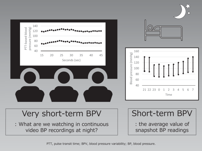
Nighttime very short-term blood pressure variability (BPV) versus nighttime short-term BPV

Full size image

Nighttime short-term blood pressure variability (BPV) and nighttime very short-term BPV

During non-rapid eye movement (NREM) sleep, there is an increase in parasympathetic activity and a reduction in cardiac sympathetic activity. By contrast, rapid eye movement (REM) sleep is a state of autonomic instability, which is dominated by remarkable fluctuations between parasympathetic and sympathetic influences. When the circadian rhythm of BP is normal, the short-term BPV in nighttime BP decreases by 10–20% of the daytime level on waking. A report using a cardiopulmonary coupling analysis showed that normotensive subjects had very-short BPV during sleep and that during NREM stage 3, deep sleep, there was a clear decrease in very-short BPV, reflecting suppressed vascular-sympathetic activity and a trend in elevated baroreflex sensitivity [3]. Nighttime very short-term BPV reflects baroreflex sensitivity, autonomic function, and respiratory movement [4]. Patients with HF have increased neurohumoral factors and arterial stiffness compared with normal subjects, and it is expected that these factors would significantly impact the increase in very short-term BPV at night. In a report by Sato et al. [2], there was a very weak but significant correlation between the value of the cardio-ankle vascular index and the coefficient of variation (CoV) for systolic BP and between the value of hemoglobin A1c and CoVs for systolic and diastolic BP. This may have been due to the possibility that the decreased arterial wall distensibility associated with elastic arteriosclerosis and autonomic neuropathy associated with abnormal glucose metabolism caused a decrease in sensitivity and dysfunction of the baroreceptor reflex system, which may have contributed to the increase in very short-term BPV. Arousal is also one of the causes of increased nighttime very short-term BPV. Arousal from sleep can occur through respiratory efforts, limb movement, and spontaneous arousal [5, 6]. Arousal induces autonomic changes, resulting in increases in nighttime BP. In a study by Davies et al., healthy subjects were assessed for very short-term BPV during each sleep stage using beat-to-beat BP monitoring [7]. When arousal occurred, the average systolic BP/diastolic BP rose 10.0/6.1 mmHg during NREM sleep and 6.0/3.7 mmHg during REM sleep, compared with control values, over the 10 s following the application of a stimulus to induce transient arousal [7]. HF patients characterized by increased sympathetic nervous system activity often suffer from sleep problems [8]. The mechanism by which nighttime very short-term BPV increases in HF patients through frequent arousals cannot be ignored.

Although some reports have shown a correlation between an increase in very short-term BPV and organ damage, there is no consensus as to whether very short-term BPV is a cause or a result of organ damage, i.e., whether it can be a therapeutic target or is merely a surrogate marker for organ damage.

Noninvasive nighttime blood pressure measurement

The BPV in nighttime BP is smaller than that in daytime BP. Therefore, if nighttime BP measurement were possible without any burden, nighttime very short-term BPV might make it easier to grasp a subject’s true intrinsic condition because extrinsic stimuli decrease at night. In healthy individuals, even stimuli during sleep that did not cause electroencephalogram arousal increased BP [7]. Cuff-based ambulatory BP machines cause appreciable arousal from sleep and therefore alter the BP that they are trying to record [9]. Although the pulse transit time-based continuous BP monitoring method [10] used in this study has the limitation of requiring frequent calibration, it may be less intrusive and may be useful for measuring nighttime very short-term BPV and short-term BPV.

Related Articles

Iron homeostasis and ferroptosis in muscle diseases and disorders: mechanisms and therapeutic prospects

The muscular system plays a critical role in the human body by governing skeletal movement, cardiovascular function, and the activities of digestive organs. Additionally, muscle tissues serve an endocrine function by secreting myogenic cytokines, thereby regulating metabolism throughout the entire body. Maintaining muscle function requires iron homeostasis. Recent studies suggest that disruptions in iron metabolism and ferroptosis, a form of iron-dependent cell death, are essential contributors to the progression of a wide range of muscle diseases and disorders, including sarcopenia, cardiomyopathy, and amyotrophic lateral sclerosis. Thus, a comprehensive overview of the mechanisms regulating iron metabolism and ferroptosis in these conditions is crucial for identifying potential therapeutic targets and developing new strategies for disease treatment and/or prevention. This review aims to summarize recent advances in understanding the molecular mechanisms underlying ferroptosis in the context of muscle injury, as well as associated muscle diseases and disorders. Moreover, we discuss potential targets within the ferroptosis pathway and possible strategies for managing muscle disorders. Finally, we shed new light on current limitations and future prospects for therapeutic interventions targeting ferroptosis.

Type 2 immunity in allergic diseases

Significant advancements have been made in understanding the cellular and molecular mechanisms of type 2 immunity in allergic diseases such as asthma, allergic rhinitis, chronic rhinosinusitis, eosinophilic esophagitis (EoE), food and drug allergies, and atopic dermatitis (AD). Type 2 immunity has evolved to protect against parasitic diseases and toxins, plays a role in the expulsion of parasites and larvae from inner tissues to the lumen and outside the body, maintains microbe-rich skin and mucosal epithelial barriers and counterbalances the type 1 immune response and its destructive effects. During the development of a type 2 immune response, an innate immune response initiates starting from epithelial cells and innate lymphoid cells (ILCs), including dendritic cells and macrophages, and translates to adaptive T and B-cell immunity, particularly IgE antibody production. Eosinophils, mast cells and basophils have effects on effector functions. Cytokines from ILC2s and CD4+ helper type 2 (Th2) cells, CD8 + T cells, and NK-T cells, along with myeloid cells, including IL-4, IL-5, IL-9, and IL-13, initiate and sustain allergic inflammation via T cell cells, eosinophils, and ILC2s; promote IgE class switching; and open the epithelial barrier. Epithelial cell activation, alarmin release and barrier dysfunction are key in the development of not only allergic diseases but also many other systemic diseases. Recent biologics targeting the pathways and effector functions of IL4/IL13, IL-5, and IgE have shown promising results for almost all ages, although some patients with severe allergic diseases do not respond to these therapies, highlighting the unmet need for a more detailed and personalized approach.

Targeting of TAMs: can we be more clever than cancer cells?

With increasing incidence and geography, cancer is one of the leading causes of death, reduced quality of life and disability worldwide. Principal progress in the development of new anticancer therapies, in improving the efficiency of immunotherapeutic tools, and in the personification of conventional therapies needs to consider cancer-specific and patient-specific programming of innate immunity. Intratumoral TAMs and their precursors, resident macrophages and monocytes, are principal regulators of tumor progression and therapy resistance. Our review summarizes the accumulated evidence for the subpopulations of TAMs and their increasing number of biomarkers, indicating their predictive value for the clinical parameters of carcinogenesis and therapy resistance, with a focus on solid cancers of non-infectious etiology. We present the state-of-the-art knowledge about the tumor-supporting functions of TAMs at all stages of tumor progression and highlight biomarkers, recently identified by single-cell and spatial analytical methods, that discriminate between tumor-promoting and tumor-inhibiting TAMs, where both subtypes express a combination of prototype M1 and M2 genes. Our review focuses on novel mechanisms involved in the crosstalk among epigenetic, signaling, transcriptional and metabolic pathways in TAMs. Particular attention has been given to the recently identified link between cancer cell metabolism and the epigenetic programming of TAMs by histone lactylation, which can be responsible for the unlimited protumoral programming of TAMs. Finally, we explain how TAMs interfere with currently used anticancer therapeutics and summarize the most advanced data from clinical trials, which we divide into four categories: inhibition of TAM survival and differentiation, inhibition of monocyte/TAM recruitment into tumors, functional reprogramming of TAMs, and genetic enhancement of macrophages.

Integrated proteogenomic characterization of ampullary adenocarcinoma

Ampullary adenocarcinoma (AMPAC) is a rare and heterogeneous malignancy. Here we performed a comprehensive proteogenomic analysis of 198 samples from Chinese AMPAC patients and duodenum patients. Genomic data illustrate that 4q loss causes fatty acid accumulation and cell proliferation. Proteomic analysis has revealed three distinct clusters (C-FAM, C-AD, C-CC), among which the most aggressive cluster, C-AD, is associated with the poorest prognosis and is characterized by focal adhesion. Immune clustering identifies three immune clusters and reveals that immune cluster M1 (macrophage infiltration cluster) and M3 (DC cell infiltration cluster), which exhibit a higher immune score compared to cluster M2 (CD4+ T-cell infiltration cluster), are associated with a poor prognosis due to the potential secretion of IL-6 by tumor cells and its consequential influence. This study provides a comprehensive proteogenomic analysis for seeking for better understanding and potential treatment of AMPAC.

Structure and function relationships of mucociliary clearance in human and rat airways

Mucociliary clearance is a vital defense mechanism of the human airways, protecting against harmful particles and infections. When this process fails, it contributes to respiratory diseases like chronic obstructive pulmonary disease (COPD) and asthma. While advances in single-cell transcriptomics have revealed the complexity of airway composition, much of what we know about how airway structure impacts clearance relies on animal studies. This limits our ability to create accurate human-based models of airway diseases. Here we show that the airways in female rats and in humans exhibit species-specific differences in the distribution of ciliated and secretory cells as well as in ciliary beat, resulting in significantly higher clearance effectiveness in humans. We further reveal that standard lab-grown cultures exhibit lower clearance effectiveness compared to human airways, and we identify the underlying structural differences. By combining diverse experiments and physics-based modeling, we establish universal benchmarks to assess human airway function, interpret preclinical models, and better understand disease-specific impairments in mucociliary clearance.

Responses

Your email address will not be published. Required fields are marked *